The urinary system is efficient in filtering the blood to remove substances that are harmful to the body. The kidneys and their associated parts all play a role in making sure that the blood is kept clean and that the body does not lose too much water or other essential nutrients. The urinary system also has other important roles in keeping the balance, or homeostasis of the blood.
The organs of the urinary system are the kidneys, ureters, urinary bladder and the urethra. The kidneys really do the work, while the other parts act as viaducts or storage for what the kidneys produce.
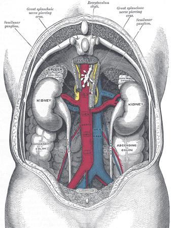
Diagram of Kidneys
The kidneys filter many liters of blood every day—approximately one-quarter of the body’s blood flows through the kidneys each minute! In addition, they regulate the blood’s volume and its chemical makeup.
The kidneys help regulate blood pressure with the production of an enzyme called rennin. When blood pressure drops, rennin causes blood vessels to constrict, forcing blood pressure to increase. The kidneys produce the hormone erythropoietin, which stimulates the production of red blood cells. The kidneys also convert Vitamin D into a form that is usable by the body.
In addition, the kidneys have four main roles:
Nitrogen containing wastes include:
Blood pH is 7.35–7.45. Alkalosis is above 7.45 and acidosis is below 7.35. These disorders are a result of excess oxygen or carbon dioxide in the blood as well as byproducts of lactic acid and fatty acid breakdown.
There are blood buffers that help prevent these changes. These buffers bind to the H+ when the pH drops and release H+ when the pH increases (bicarbonate buffer system). The kidneys get rid of acids that are generated during metabolism by excreting or reabsorbing bicarbonate as needed to effect change in the pH.
The third and fourth roles of the kidneys—water and electrolyte balance—are closely linked.
Water makes up 50 to 60 percent of body weight depending on gender. On average, women tend to have more fat than men, and fat has less water than muscle. Two-thirds of water is found in cells, while the remaining third is found extracellularly, meaning in interstitial fluid, plasma, cerebral spinal fluid, humors of the eye, and the like.
The re-absorption of water and electrolytes is regulated mostly by two hormones: anti-diuretic hormone (ADH) and aldosterone (controlled by rennin). If there is high blood loss and blood pressure drops, the filtrate drops. Reacting to this change, osmoreceptors in the hypothalamus (in the brain) release ADH. This allows for the absorption of more water in order to increase blood volume and pressure; therefore only a small amount of concentrated urine is produced. If there is too little Na+ or other ions in the blood, the blood becomes diluted, which causes edema or circulatory collapse. Aldosterone regulates the re-absorption of Na+. As it is reabsorbed, water passively follows Na+.
In review, the kidneys function to regulate several processes:
Additionally, the kidneys:
The kidneys are located between T12 and L3 of the spine in the mid- to lower-back and are protected by the ribs. The right kidney is a little lower than the left due to the location of the liver.
Each kidney is shaped like a kidney bean. The indention is called the hilus. Kidneys are encased in a protective sheath called the renal capsule. The renal capsule and some adipose tissue hold the kidney against the muscles of the trunk wall.
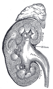
Three Kidney Sections
A kidney opened lengthwise reveals three regions:
The pyramids are arranged with the base of the pyramid toward the cortex and the apex towards the inner part of the kidney. At the end of the pyramids are collection vessels called calyces. The calyces drain into the renal pelvis, a cavity that is continuous with the ureter. The pyramids are separated by renal columns, which are structures that are similar in makeup to the cortex.
Since so much blood passes through the kidneys, they have a great blood supply. The renal artery comes off of the abdominal aorta that goes to the kidneys. From there it segments into the segmental artery, to the lobular artery, into the interlobular arteries, and then to the cortex.
The major structural unit of the kidney is the nephron. In each kidney there are millions of nephrons producing urine. A nephron has two main parts: the glomerulus and the renal tubule.
The glomerulus is essentially a capillary bed. The blood goes into the glomerulus through the afferent arteriole and leaves via the efferent arteriole. At one end of the renal tubule there is a capsule (Bowman’s) that cups around the glomerulus, forming a porous membrane. Extending from the Bowman’s capsule, the renal tubule becomes very convoluted and is surrounded by blood vessels called peritubular capillaries. The renal tubule has three parts: the proximal tubule, the Loop of Henle, and the distal tubule. The proximal tubule ascends down to the Loop of Henle and then ascends back up in the distal tubule.
The major functional part of the kidneys is the
B is the correct answer. The hilus is a depression where vessels, nerves, or ducts enter the kidney. The renal cortex contains the glomeruli and the convoluted tubules. Renal tubules are small structures that filter blood and produce urine.
Filtration occurs between the glomerulus and the Bowman’s capsule by high pressure in the glomerulus. This pressure basically pushes everything that is small enough—water, ions, vitamins, amino acids, and wastes—into the Bowman’s capsule. However, if blood pressure is too low, there is not enough pressure and filtration will not occur. As the filtrate runs through the renal tubule, most of it is reabsorbed into the peritubular capillaries.
The peritubular capillaries arise from the efferent arterioles. The capillaries envelop the renal tubules before draining into the venules. The peritubular capillaries are porous and have low pressure, yet have a high absorption of solutes and water during reabsorption. The solutes and water are then returned to the bloodstream.
The body wants water, glucose, amino acids, and ions, so the body reabsorbs almost everything that was pushed out. Reabsorption begins as soon as the filtrate enters the proximal convoluted tubule. Unlike the filtration, which was simple passive transport, reabsorption often uses active transport. There are carriers for the substances that the body wants to keep, like glucose and amino acids. These usually are all removed and are not found in urine. However, there are no carriers for the substances we don’t want to keep—like creatine and uric acid. Ions are reabsorbed as necessary.
It is while the filtrate is passing through the nephron that urine is formed. Generally, the kidneys filter 150-180 liters of plasma in 24 hours, yet produce only 1-2 liters of urine.
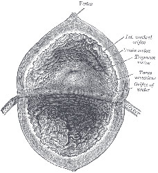
Diagram of Bladder
Under normal conditions, urine is sterile, yet slightly acidic (~6.0 pH). Coloration ranges from clear to dark yellow, the latter due to the urochrome pigment from the breakdown of hemoglobin. Urine can contain Na+, K+, urea, uric acid, creatinine, ammonia, ions, and vitamins.
Urine in the kidneys needs to leave the body. This is where the other parts of the urinary system come into play.
The ureters are tubes that go from the hilus of the kidney to the urinary bladder. Ureters are smooth muscle and use peristalsis.
The urinary bladder is a smooth collapsible sac that holds urine. This bladder expands as it fills.
The urethra is a tube that carries the urine from the bladder out of the body. The urethra also operates through peristalsis. It has an internal sphincter that stays closed when urine is not being expelled. Urethra size depends on gender: it is much shorter in females (3–4 cm) compared to males (20 cm).