In this lesson, you will review the structures and functions of plant cells.
Plant cells share many characteristics with animal cells. Plant cells use cellular respiration for energy, as reviewed earlier. They also have a plasma membrane, cytoplasm, a nucleus and nucleolus surrounded by a nuclear membrane, mitochondria, rough and smooth endoplasmic reticula, Golgi bodies, peroxisomes, microtubules, and other structures. However, plant cells have some unique structures, and carry out some unique, and vital, processes.
The most distinguishing characteristic of plant cells is their rigid cell wall that surrounds the inner plasma membrane. A plant cell wall is made from cellulose, which is made from glucose. Thin chains of these glucose molecules are joined as microfibrils, which give cellulose crystal-like properties. The microfibrils are wound together into threads, which themselves are wound into cables. Cellulose is the most abundant substance in plants, but lignin is a close second.
Lignin, is a glucose-based polymer that adds further rigidity and strength to the cell wall. Lignin is the major material in wood.
Plants need strong, rigid cell walls for two important reasons. First, the cell walls help support the plant; for example, keeping its stem upright. Second, water drawn up through the plant via the roots is under pressure, and strong cell walls are required to maintain cell shape, and prevent cell explosion, during water intake.
Vacuoles are, in some ways, similar to vesicles in animal cells. However, plant cell vacuoles are usually far larger than vesicles (taking up 90 percent of the space in some plant cells). Vacuoles are membrane-bound sacs filled with liquid called cell sap, which is mostly water. In some plants, the cells’ vacuoles hold pigments that give the plant its color, such as the red pigment in cherries.


What are the two main substances that give rigidity to the cell wall?
The correct answer is B. Cellulose forms microfibril chains that, along with lignin, add rigidity to plant cell walls. Microtubules are cytoskeletal structures in animal cells, so A is not correct. Thylakoids are pigment-carrying parts of chloroplasts, so C is incorrect. Grana are stacks of thylakoids, so D is also not correct.
The grana that occur in chloroplasts are primarily made up of
The correct answer is C. Grana are the stacks of pigment-rich thylakoids that occur in the chloroplasts. A chloroplast is itself a plastid, a type of organelle, but its constituents are not plastids, so A is not correct. The stroma is the liquid that fills the area around the grana in the chloroplasts, so B is not correct. Carotenoid pigments may be incorporated into the thylakoids that make up the grana, but the grana are not made up of these pigments, so D is not correct.
Plants are autotrophs because they make their own food via the process of photosynthesis. Photosynthesisis a complex chemical process in which the energy in sunlight is used to convert carbon dioxide and water into a form of glucose a plant can use as food, with oxygen as a byproduct. Photosynthesis takes place in the chloroplasts, particularly in the thylakoids.
Of course, plants take in water through their roots, and the water is drawn upward to every cell in the plant. In most plants, the underside of the leaf contains small, pore-like opening called stomates that take in air.
This is the chemical formula for photosynthesis:
CO2 + H2O → light energy → (CH2O) + O2
CH2O is the generic formula for carbohydrates used as food by plants.
Different plant pigments absorb different wavelengths, or colors, of light. Chlorophyll, reflects the color green to give most plants their green color. Chlorophyll absorbs and utilizes wavelengths of light in the violet-blue-red range of the spectrum. Other plant pigments, including carotenoids and phycobilins, absorb other spectra of light to provide a photosystem of complementary that further utilize wavelengths of light outside the operative range of chlorophyll. These accessory pigments are most visible in the autumn as the leaves lose their chlorophyll (green color) to expose the underlying colors of the remaining pigments.
A photosystem includes chlorophyll and other pigments that are embedded in thylakoids. One chlorophyll a molecule per photosystem absorbs one photon of light. The energy boosts one of the chlorophyll a electrons to a higher level. It moves to an acceptor molecule, and an electron flow is initiated. The chlorophyll a molecule is thus oxidized and has a positive charge. In photosystem I, the activated chlorophyll a molecule is designated P 700 (P for pigment; 700 for the optimum wavelength absorbed). In photosystem II, chlorophyll a is P 680.
P 680 is excited by a photon of energy from incoming light, and its excited electron is transferred to an acceptor molecule. The P 680 is then able to replenish its electrons from a water molecule it splits in a process known as photolysis. The electrons cascade along an electron transport chain, forming ATP in the process. The term for this process is photophosphorylation.
In photosystem I, electrons from P 700 are passed downhill until they reach the coenzyme NADP, which is reduced to form NADPH2. NADPH2 provides energy for the synthesis of fuel for the cell.
Thus, in the light reaction, electrons flow continuously from water to photosystem II, and from there to photosystem I and to NADP.
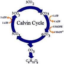
Despite its name, the dark reaction often takes place in the presence of light, though light is not required. In this second stage of photosynthesis, carbon from carbon dioxide is chemically reduced. The process occurs in the stroma, and this carbon fixation process is referred to as the Calvin cycle after the scientist Melvin Calvin, who first described it.
The Calvin cycle begins and ends with a five-carbon sugar that has two phosphate groups attached to it (ribulose 1.5-biphosphate, or RuBP). Carbon dioxide is attached to the RuBP, which then splits to form two molecules of PGA (3-phosphoglycerate). The cycle must repeat six times for the final product, glyceraldehyde 3-phosphate, to result.
6 CO2 + 12 NADPH2 + 18 ATP ⇒ 1 glucose + 12 NADP + 18 ADP + 18P i + 6 H2O
C4 plants do not use the Calvin cycle to fix carbon. Instead of using the 3-carbon PGA molecule, they use the 4-carbon oxaloacetate. The oxaloacetate is reduced to malate or aspartate, and it is then decarboxylated to yield CO2 and pyruvate. The CO2 then enters the normal Calvin cycle, while the pyruvate reacts with ATP to form phosphoenolpyruvate (PEP). This rather roundabout and energy-consuming process may seem inefficient at first, but it has its advantages, especially under conditions in which photorespiration occurs.
Photorespiration is a process in plants in which oxygen is consumed and carbon dioxide is released in the presence of light. Photorespiration always accompanies the Calvin cycle and occurs most often during periods of intense light or heat or reduced water availability. The problem with photorespiration, which occurs in the peroxisomes, is that it produces neither ATP nor NADH2. What it does produce is lots of carbon dioxide—up to 50 percent of the carbon fixed during photosynthesis may be reoxidized to CO2 during this process and then released through leaf stomates. This appears to be an inefficient use of energy.
C4 plants, however, can refix this excess CO2 by cycling it into the C4 system, so it is not wasted. The PEP involved in C4 photosynthesis has a greater affinity for carbon dioxide than RuBp, so more CO2 is taken up and used. In fact, the photosynthetic rate in plants that use the C4 pathway (corn, sugarcane, sorghum, etc.) is three to four times that of plants using the Calvin cycle.
In the designation P 680, the subscript number represents
The correct answer is B. The subscript number of the pigment indicates the wavelength of light it best absorbs. The subscript number does not indicate the number of atoms in the molecule of pigment, so A is not correct. The number of glucose molecules produced in the Calvin cycle varies and is not dependent on the type of pigment, so C is incorrect. The number of carbon dioxide molecules fixed during photosynthesis depends on conditions and is not indicated by the subscript wavelength number designating the pigment; thus, D is not correct.
The C4 photosynthetic pathway is more efficient than the Calvin cycle in that it
The correct answer is D. In the C4 cycle, carbon molecules are reduced and the carbon dioxide that results is cycled into the Calvin cycle instead of being released; so this is a more efficient use of carbon in photosynthesis. All cell functions require the energy of ATP, which has nothing to do with the double processing of the more efficient C4 cycle, so A is not correct. B is not correct because PEP is an intermediate material made during C4 photosynthesis and does not explain why this type of photosynthesis uses carbon more efficiently. The C4 cycle does not use fewer carbon dioxide molecules; it just uses the ones it has completely instead of losing them; so C is not correct.
In photosynthesis, an electron cascade results from photolysis in which
The correct answer is C. In photolysis, a pigment traps an excited electron, whose energy it uses to cleave a water molecule into its component hydrogen and oxygen. A photon is not itself transferred among molecules, but the photon-activated electron is; so A is not correct. B is not correct because carbon fixation is a process separate from photolysis, which involves the splitting of water molecules. Photolysis does not involve the oxidation and release of carbon dioxide; reoxidation of carbon dioxide occurs in photorespiration. So D is not correct.
All cells reproduce when a parent cell splits in two to form two genetically identical cells, called daughter cells. Reproduction involving only one parent that produces genetically identical offspring is a form of asexual reproduction. Mitosis is the duplication of the cell’s genetic material. Cell division, or cytokinesis, is the actual splitting off of the daughter cells.
Mitosis is part of the cell cycle, or the life cycle of the cell. The cell cycle is the normal sequence of cell development, functioning, and division. The cell cycle has two primary phases. During the first phase, the cell carries out its normal functions. This stage is called interphase. This part of the cell’s life is very busy during this time—carrying out metabolism, transport, respiration, and so on. During interphase, a cell grows to its normal size, which is generally about twice the size it was when it was first produced. It is also during interphase that the cell readies itself for mitosis. The second broad phase of the cell cycle occurs when the cell divides via mitosis and cytokinesis. Each cell cycle phase may take hours, days, or even longer, depending on the cell.
Interphase is usually the longest period in a cell’s life. It is such a busy period in the life of the cell that it is divided into several phases. Once cytokinesis is complete, the newly formed cell increases to its normal size, produces significant amounts of ATP, and synthesizes enzymes and proteins. This occurs during the G1 (Gap 1) phase of interphase. The duration of G1 phase differs depending on the type of cell. Some cells do not proceed to the preparatory cell division phases that follow, but may continue indefinitely (or for their entire lives) in the G1 phase.
Eukaryotic cells contain far more chromosomes and DNA than prokaryotes. So the process of ensuring that its genetic material is copied exactly is a more complex process in eukaryotes. The discussion that follows refers to mitosis in eukaryotic cells.
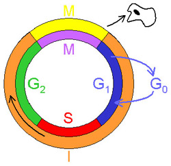
Cell cycle
In G1 phase, the cell’s chromosomes are made up of single strands of chromatin, the DNA, histones, and other materials that make up chromosomes. When the cell is ready to make preparations for mitosis, it enters S phase (Synthesizing phase), during which it synthesizes, or replicates, its DNA. This stage usually lasts several hours and produces a second, identical set of cell chromosomes.
The cell then enters G2 (Gap 2) phase, during which time the cell prepares the necessary structures needed for mitosis and cytokinesis. The cell still retains its duplicate sets of chromosomes at this stage. This is another hectic period in a cell’s life. The cell in G2 phase may double in size. It also is extremely active biochemically as it doubles the number of its organelles, ribosomes, enzymes, and other substances and structures prior to mitosis. Mitosis itself is divided into four substages.
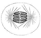
When the cell enters prophase, by far the longest mitotic phase, it has already replicated its chromosomes. As prophase begins, the cell’s chromosomes condense and become tightly coiled. It is at this stage that the chromosomes become visible under a microscope. Each chromosome now consists of two identical, or duplicated, chromatids — the identical strands of the chromosomes. (A chromatidis the single strand of a duplicated chromosome.) The chromatids are pressed closely together lengthwise and joined in the center by a centromere, a specialized region of the chromosome to which a spindle will be attached. At either side of the cell’s nucleus, at the outside of the nuclear membrane, is a pair of centrioles, organelles that help organize the spindle during mitosis. In each pair, one centriole is mature and the other has just formed.
The cell’s shape then changes and becomes more spherical. The cell’s cytoplasm becomes more viscous. During prophase, the all-important spindle begins to form. The spindle is made up of the proteins actin and tubulin. Late in G2 phase, tubulin makes up about 10 percent of the cell’s protein and, in prophase, this protein begins to assemble itself into the microtubules of the spindle. The materials that make up these microtubules are called spindle fibers. Centromeric spindle fibers are attached to a chromosome’s centromere at one end and to a pole of the spindle, and to a centriole, at the other end. Fibers that comprise the aster radiate outward from each centriole.
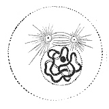
While the spindle is forming, the nuclear envelope begins to disintegrate. Toward the end of prophase, the nuclear membrane no longer exists and the condensed chromatids are no longer separated from the cell cytoplasm.
As prophase nears its end, the now equally sized centrioles have migrated to opposite sides of the cell. The fully formed, three-dimensional spindle now looks like a see-through football made of microtubules. The spindle has a full complement of centromeric fibers that attach to the kinetichores of the centromere of each chromatid; aster rays radiate outward from the centriole; and polar microtubules appear to run the length of the spindle.
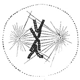
At the onset of metaphase, all structures are in place and ready for mitosis. At metaphase, the chromosomes consist of identical pairs of chromatids cinched together by a centromere. Spindle fibers are attached to each centromere, and the spindle fibers maneuver the chromosomes to the center of the cell, or the equator of the spindle. There the chromosomes—more precisely, their centromeres—line up in the center of the spindle along a plane called the metaphase plate.
At the beginning of anaphase, the centromeres begin to separate, pulled apart by the spindle fibers attached to them. One set of centromeres is pulled by the spindle fibers to one pole of the cell; the other set is pulled to the opposite pole. As the centromeres are pulled apart, the chromatids pairs are, likewise, separated and move to opposite poles of the cell. When the chromatids are separated, they are again referred to as chromosomes. By the end of anaphase, two identical sets of chromosomes occur on opposite poles of the spindle. Anaphase is the briefest stage of mitosis.
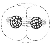
With the identical sets of chromosomes at the poles of the spindle, telophase begins. Early in telophase, the spindle begins to disappear, and the microtubules revert to tubulin proteins. As telophase continues, a nuclear envelope begins to form around each set of chromosomes. By late telophase, a nuclear envelope completely surrounds each chromosome set. By this time, the chromosomes have de-condensed and become more diffuse. Nucleoli reappear, and a new centriole often forms. During the end stage of telophase, the plasma membrane begins to pinch closed at its center.
Cytokinesis is the division of the cell’s cytoplasm. The plasma membrane continues to pinch inward at the midline of the cell, which is always along the metaphase plate, where the equator of the spindle had been situated. Actin microtubules just beneath the plasma membrane are responsible for guiding the creation of the furrow and the final cleavage of the cell. Eventually the two sides of the membrane touch, and the cytoplasm effectively divides into two cells.
There are some ways that cytokinesis differs in animal and plant cells. In animal cells, actin filaments near the plasma membrane create a groove that contracts along the site of the metaphase plate until the daughter cells are separated. Plant cells, however, have rigid cell walls that cannot gracefully curve inward toward a wasp-waisted separation of the daughter cells. Instead, the cytoplasm of dividing plant cells is separated by the formation of a cell plate, which forms from a series of vesicles produced by the Golgi bodies. The vesicles start to fuse from the center outward to create the flat, membrane-bound cell plate. When the cell plate touches the plasma membrane, the two daughter cells are separated. The membranous space of the cell plate fills up with pectins and, eventually, each daughter cell constructs a new cell wall on her side of the cell plate by layering cellulose along the membrane.
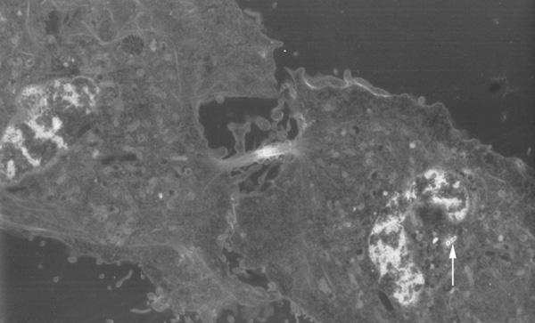
Cytokinesis electron micrograph
The process of mitosis is intended to
The correct answer is D. The primary purpose of the mitotic process is to assure the correct replication and subsequent division of a cell’s chromosomes. Two daughter cells are the outcome of cell division, not the primary purpose of mitosis. So A is not correct. Cytokinesis is the division of the cell cytoplasm to the daughter cells, so B is incorrect. Mitosis begins in interphase and extends beyond it, so C is not correct.
The correct answer is A. The two Gap phases and the S phase in which chromosomes are replicated all occur in interphase. Metaphase sees the centromeres, attached to spindle fibers, aligned along the metaphase plate, so B is incorrect. In anaphase, the spindle pulls the paired chromosomes apart, so C is not correct. Telophase occurs when the two daughter cells begin to form and the spindle starts to disintegrate, so D is not correct.
The correct answer is C. A chromatid is a single strand of a duplicated chromosome. The centriole is an organelle that occurs on either side of the nuclear membrane. Centrioles help organize the spindle, so A is not correct. A centromere holds the paired chromatids together at the center, so B is not correct. Chromatin is the single strands of DNA that make up the cell’s chromosomes, so D is not correct.
The correct answer is B. Spindle fibers attached to the centromeres of each chromosome pair pull the pair apart and each chromosome migrates to one pole of the cell. Asters occur at the poles of the cell and are not attached to the chromosomes, so A is incorrect. Spindle fibers are not attached to the metaphase plate, but to the centromeres; C is not correct. Microtubules make up the spindle but do not cleave the cell, so D is not correct.
The correct answer is A. A cell plate grows between the two daughter cells, eventually attaching to the cell membrane and dividing the two cells. B is not correct because it is not the Golgi bodies that create the cell plate, though they create the vesicles that begin to form the plate. The cell plate is made of membranous vesicles that eventually fill with pectin. They do not contain lignin, so C is incorrect. Cellulose gives a cell wall some of its rigidity. It is not part of a plant cell’s membranes, so D is not correct.
The correct answer is B. The pinching inward of the membrane in animal cells or the creation of a cell plate in plant cells separates the cytoplasm in the two daughter cells. The cell membrane in an animal cell pinches inward, and no new membrane forms, so A in not correct. A new nuclear envelope does form in the daughter cells, but this is not part of cytokinesis, so C is incorrect. The plasma membrane does not rupture, but creates a groove or cleavage in the middle of the cell as the daughter cells form, so D is not correct.