In this lesson, you will review how DNA technology is used in medicine, forensic science, and other fields.
Studying a particular gene is difficult. The genes constitute only a small fraction of the total amount of DNA in a cell. On the other hand, a single molecule of DNA holds many different genes. A single gene might be only 1/100,000th of the molecule in which it resides. Making things even more difficult, DNA molecules are very homogenous over their length, since they consist of sequences of only four different nucleotides. DNA cloning makes it possible for the biologist to make multiple copies of a single gene.
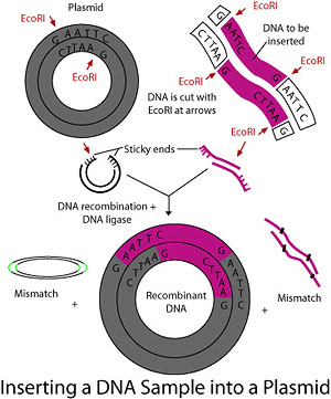
One technique for cloning a gene uses the reproductive machinery of bacteria as “factories” to make copies of the gene in a process known as bacterial transformation. Recall that a plasmid is a small bit of DNA that is separate from the bacterial chromosome. Plasmids are copied along with the chromosome when the bacterial cell divides. DNA cloning requires removing plasmids from bacterial cells, cutting the plasmid at one point using a restriction enzyme, inserting the desired gene into the plasmid, rejoining the ends with DNA ligase forming recombinant DNA, then re-inserting the plasmid back into a bacterial cell.
Unfortunately, it is not quite that simple. When the human chromosome is cut up into genes using a restriction enzyme, many different genes will likely be present. All of these genes will get inserted into bacterial plasmids. When the bacteria reproduce on growth medium, many different colonies will grow and only some of these colonies will contain the gene of interest. In most cases, however, it is relatively easy to identify the colonies with the new gene expressed. For example, if the researcher is studying a gene that confers ampicillin resistance, then ampicillin can be added to the growth medium. Then, only colonies with that gene will grow.
Often, however, the colonies with the gene of interest cannot be easily distinguished from the others. In that case, nucleic acid hybridization can be used to identify the colonies with the gene. In this process, a short single strand of nucleic acid (DNA or RNA) containing a base sequence known to be complementary to a sequence on the target gene is “tagged” with a radioactive isotope or a fluorescent molecule. Then a piece of filter paper is applied to the colonies on the growth medium. Some cells are picked up and transferred to the paper. The filter is treated to denature the DNA, and then a solution containing the tagged DNA probes is applied to the paper. The excess solution is washed away and the filter paper is laid on a piece of photograph film. Either the radioactive or fluorescent tags expose the film. The film can then be compared to the still living colonies on the growth medium to identify which contain the target gene.
Before cloning can be done, both the plasmid and the chromosome must be cut. The plasmid must be cut to allow insertion of the gene, while the chromosome must be cut to isolate the gene of interest. The process of cutting the DNA into pieces is called restriction digestion. The cutting is done with special enzymes referred to as restriction endonucleases, restriction enzymes, or REs. These special enzymes recognize specific nucleotide sequences in the DNA molecule wherever that sequence occurs.
The process starts when the restriction enzyme is mixed with a sample of the DNA. However, REs are fragile and must be treated with care. Like all proteins, they denature (break hydrogen bonds) when heated. REs are kept frozen until they are needed. All of the ingredients in a restriction digest are kept cold until it is time for the reaction to begin. In addition, salinity and pH must be controlled. Chemical buffers are used to assure that the concentration of various substances, such as NaCl and MgCl2, remains close to the optimum value. When everything is ready, the temperature of the reaction vessel is carefully raised to the appropriate temperature, which is different for each RE and gene. When the digest has run for the appropriate amount of time (usually one hour), the reaction tube is immediately put on ice to prevent degradation of the DNA fragments. The fragments are then separated by gel electrophoresis to visualize the fragments and possibly purify them for further experiments.
In recombinant DNA experiments, substance X is used to cut pieces of DNA, and substance Y joins these segments to form recombinant DNA. Which of the following choices correctly pairs these two substances?
A is the correct answer. Choices B and C are incorrect because a plasmid is the structure in a bacterial cell into which the DNA is inserted. Choice D has the correct two substances, but in the wrong order.
Gel electrophoresis is a technique used by scientists to separate molecules based on physical characteristics, such as size, shape, or electrical charge. The gel used is a polymer with controlled porosity. Small molecules use a gel made of polyacrylamide. Larger molecules, such as genes, large proteins, and nucleic acid sequences longer than a few hundred bases, use agarose. Agarose is a seaweed extract similar to the growth medium agar. In both cases, the gel forms a gelatinous semi-solid and porous material that looks and feels similar to food gelatin.
The gel is supported between two glass plates. Electrodes are placed on the top and bottom edges of the gel. In solution, nucleic acids and other macromolecules have ionic characteristics, usually from the phosphate group on the sugar-phosphate backbone. The solutions containing the molecules to be analyzed are added to small “wells” at the top and an electric field is applied to the two electrodes. The negatively-charged molecules are attracted to the bottom plate and repelled by the top plate, so they start to move through the gel. However, because of the limited and controlled porosity of the gel, the molecules move relatively slowly. Small, less tangled molecules move more quickly, while larger or more tangled molecules move more slowly.
Proteins can have both positive and negative charges. So proteins are usually denatured by mild heating in the presence of a detergent that coats the proteins with a negative charge. Generally, the amount of detergent bound to the protein is relative to the size of the protein, so that the resulting denatured proteins have an overall negative charge, and all the proteins have a similar charge-to-mass ratio. After denaturing, the proteins also have a rod-like shape similar to DNA, so they move through the gel in a similar way.
After electrophoresis, the molecules in the gel can be stained to make them visible. However, there may be thousands of different proteins or nucleic acid strands. If the gel is stained to make them visible, each lane may appear as a solid bar. So a technique similar to the one used to identify bacterial colonies can be used to highlight only the molecules of interest. For example, the gel can be treated with a DNA probe that contains a fluorescent dye with nucleotide sequences complementary to known sequences in the target molecules.
Typically, several mixtures are placed in adjacent wells in the gel. They then migrate toward the other electrode, forming parallel stripes in the gel. Each lane exhibits separation of the components from the original mixture as one or more distinct bands, one band per component. Bands in different lanes that end up the same distance from the top contain molecules that passed through the gel with the same speed, which means they are approximately the same size. A gel can also be “calibrated” by adding a mixture of known molecules to one of the wells. Then, unknown bands can be compared to the known bands. Sometimes they can be identified. If they do not match a known molecule, then the size of the molecule can be estimated by comparison with known sizes.
Which of the following describes a nucleic acid probe?
D is the correct answer. Nucleotide probes are used to locate specific nucleotide sequences. Choice A is a description of the function of the enzyme ligase. Choice B is a description of a process. Choice C is a description of a restriction enzyme.
Southern blotting (named after its developer, British biologist Edward Southern at Edinburgh University during the 1970s) is a method in molecular biology of enhancing the result of an agarose gel electrophoresis.
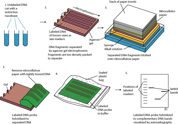
Step 2: The mixtures are separated by gel electrophoresis. The lanes of DNA strands are too densely packed to see individual molecular bands.
Step 3: Special nitrocellulose paper is placed on the gel and held in place by a stack of paper towels. The strands of DNA stick to the nitrocellulose paper.
Step 4: The paper blot is removed from the gel and placed in a plastic bag containing a radioactive probe.
Step 5: The paper blot is photographed with autoradiography. Individual molecular bands are now obvious.
The name Southern blotting has led to the somewhat whimsical naming of other techniques. Northern blotting is identical except used for RNA instead of DNA. Western blotting uses an antibody instead of a radioactive tag.
A bacterial plasmid with a new DNA segment inserted is called recombinant DNA. It is also possible to splice different segments of linear DNA together. Combining DNA from different sources has the potential to produce new, novel, and useful DNA molecules. DNA ligation is facilitated by an enzyme, T4 DNA ligase, which is derived from the T4 bacteriophage. This enzyme will easily ligate DNA fragments having overhanging, “sticky” ends. T4 DNA ligase has more difficulty with fragments having blunt ends.
DNA fingerprinting is an enormously useful tool in criminal science, but it is also useful in genealogy. The process begins with a sample containing cells, such as blood, semen, saliva, hair, or bits of skin. First, the DNA is extracted from the cells. Next, restriction fragment length polymorphism (RFLP) analysis is performed by using a restriction enzyme to cut the DNA into fragments that are separated into bands during agarose gel electrophoresis. Next, the bands of DNA are identified by Southern blotting. The visible pattern of bands on the film produced by Southern blotting (or a similar technique) is commonly called a DNA fingerprint.
Another method for genetic fingerprinting—amplified fragment length polymorphism (AFLP)—has recently been developed. The differences between RFLP and AFLP analysis include more rounds of amplification and specially made primers. AFLP analysis is now highly automated and allows for easy creation of phylogenetic trees based on comparing individual samples of DNA.
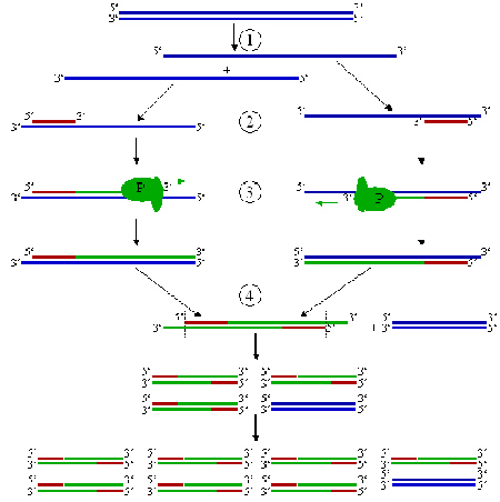
The most common, powerful, and widely accepted method for producing DNA fingerprints in criminal cases is the polymerase chain reaction (PCR). PCR involves the amplification of specific regions of DNA that are known to be highly variable from one individual to another. The amplification process allows analysis of very small amounts of material, and the outcome is highly discriminating, with the chance of a random match being in the one-in-a-billion percentile. PCR is by far the most common method for presenting DNA evidence in a criminal trial or other forensic context.
An example of the PCR cycle follows these steps:
Step 1: Denaturing at 96 degrees C.
Step 2: Annealing at 68 degrees C.
Step 3: Elongation at 72 degrees C (P = polymerase)
Step 4: First cycle is complete, resulting in two DNA strands as the templates for the next cycle. For each new cycle, the amount of DNA duplication is doubled.
Archaeologists excavated a human femur and succeeded in extracting a tiny amount of DNA from the bone. Which of the following would be the best first step in analyzing the DNA found?
C is the correct answer. The polymerase chain reaction amplifies the DNA segments. Choice A is incorrect because there may be insufficient DNA to analyze. Choice B is incorrect because fragmenting the small amount of DNA would not be productive. Choice D is useful only for identifying particular sequences.
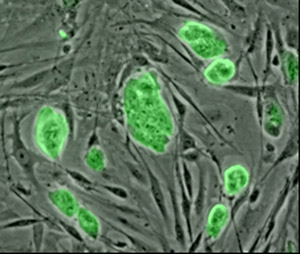
Stem cell research is one of the most promising and, at the same time, one of the most controversial areas of research in biotechnology. There are three main classes of stem cells.
Totipotent cells can develop into any other kind of cell. The zygote created by human fertilization is a totipotent cell. It can develop into any other kind of cell, including extra-embryonic cells, such as placental tissues. Many or most plant cells are totipotent. A cutting from a stem of a rose bush can be planted and will grow into a complete plant with roots, stems, leaves, and flowers.
Pluripotent cells are more limited. Embryonic stem cells are cells taken from the inner layer of the blastocyst. They can develop into any kind of human cell, but they cannot develop into placental tissue. Pluripotent stem cell research shows promise in several areas, such as cell specialization, diseases, and pharmaceutical drug testing.
Human blood cells develop from cells in the bone marrow that are said to be multipotent. Multipotent cells are even more limited. They can develop into any kind of blood cell, including red blood cells, white blood cells, and platelets. Yet, they cannot develop into muscle cells. Such terminally differentiated cells are thought to be permanently committed to a specific function.
Sources of stem cells include: cord blood from placenta and umbilical cord after birth; stem cells in all adults and young adults; and undifferentiated inner mass cells of a blastocyst.