Over the next four lessons, you will review these human body systems: nervous, endocrine, digestive, and circulatory.
 The nervous system controls and coordinates essential functions of the human body. This system receives and relays information about activities within the body and monitors and responds to internal and external changes.
Cells that carry messages throughout the nervous system are called neurons. These messages are called nerve impulses. Neurons are classified into three types according to the directions in which the impulses move.
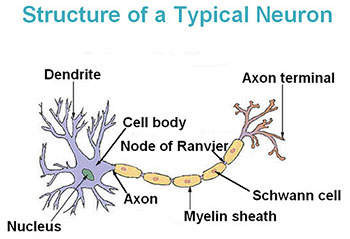
The largest part of the neuron is the cell body. The cell body contains the nucleus and most of the cytoplasm; therefore, most of the cell’s metabolic activities occur here. Spreading out from the cell body are branched extensions called dendrites. They carry impulses toward the cell body. The long fiber that carries impulses away from the cell body is the axon, which ends in a series of swellings called axon terminals.
Bundles of neurons are called nerves. The nerves contain a large number of independent communications channels. They are also composed of many supporting cells that form nervous tissue. Schwann cells wrap around axons forming a fatty, insulating covering called a myelin sheath. There are small gaps, called nodes, between the myelin sheaths. As an impulse moves down an axon, it will jump from node to node. This increases the speed of the impulse.
A nerve impulse is a flow of electrical charges along the cell membrane of a neuron. The strength of an impulse must reach a minimum level of a stimulus to activate a neuron is the threshold.
At the axon terminals, the neuron may make contact with dendrites of another neuron, with a receptor or an effector. Receptors are special sensory neurons in sense organs that receive stimuli from the external environment. Effectors are muscles or glands that bring about a coordinated response. The space between neurons where an impulse is passed from one cell to another is called a synapse. Tiny vesicles located in the axon terminal are filled with chemicals called neurotransmitters. The vesicles are stimulated to secrete the neurotransmitters into the synapse where they are received by the dendrite or receptor of the next neuron to continue the transmission of the nerve impulse.
Which type of neuron is responsible for transmitting impulses to the central nervous system?
A is the correct choice, because sensory neurons carry signals from sense organs to the brain and spinal cord. Interneurons connect sensory and motor neurons and carry impulses between them. Receptor neurons are special sensory neurons in sense organs that receive stimuli from the external environment. Motor neurons carry impulses from the brain and spinal cord to muscles or glands.
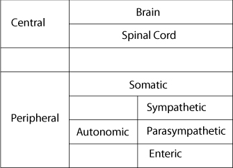
Nervous systems divisions
The human nervous system is divided into two major divisions: the central nervous system and the peripheral nervous system. The central nervous system serves as the control center of the body. It consists of the brain and the spinal cord. Its functions are relaying messages, processing information, and comparing and analyzing information. The peripheral nervous system consists of everything but the brain and spinal cord, including the cranial nerves, and nerves of the neck, chest, lower back, and pelvis.
The brain contains approximately 35 billion neurons and has a mass of 3 pounds. It is wrapped in three layers of connective tissue known as meninges. The innermost layer, the pia mater, is bound to the brain. The middle layer is the arachnoid layer. In between these layers, a space filled with cerebrospinal fluid acts as a shock absorber. The outermost layer is the dura mater. In order for the brain to function, it must have a constant supply of food and oxygen.
Brain Lobes
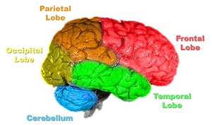
The largest part of the brain is the cerebrum. It is responsible for all of the voluntary activities of the body. It is also the sight of intelligence, learning, and judgment. The cerebrum is divided into two hemispheres, right and left, by a deep groove known as the corpus callosum. The surface of the cerebrum has numerous folds. Each hemisphere is divided into regions called lobes, which are named for the skull bones that cover them.
The right hemisphere is associated with creativity and artistic ability. The left hemisphere is associated with analytical and mathematical ability.
The cerebrum consists of two surfaces. Much of the activity occurs in the cerebral cortex, which is composed of gray matter. The gray matter contains densely packed nerve cells. The other surface of the cerebrum is the cerebral medulla. The cerebral medulla is composed of white matter, which contains bundles of myelinated axons.
The cerebellum is the second largest part of the brain and is located at the back of the skull. It controls balance, posture, and voluntary muscle contractions. The thalamus and hypothalamus are found in the part of the brain between the brain stem and the cerebrum. The egg-shaped thalamus is the main site of sensory processing. Most of the sensory nerves converge on the thalamus. Below the thalamus is the hypothalamus. It is a slender thread of tissue that controls hunger, thirst, fatigue, anger, and body temperature. It also directs the secretions of the pituitary gland.
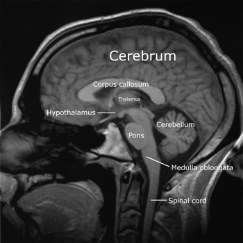
The spinal cord is continuous with the brain and emerges from the opening at the base of the skull. It stretches downward for approximately 42 to 45 centimeters. It ends just below the ribs. Like the brain, it is protected by bone, meninges and cerebrospinal fluid. The spinal cord carries impulses to and from the brain and regulates reflexes. Thirty-one pairs of spinal nerves originate in the spinal cord and branch out to the body.
Coordination and balance occur principally in the
The correct choice is B, because the cerebellum controls posture, balance, and voluntary muscle contractions. The spinal cord carries impulses to and from the brain and regulates reflexes. The cerebrum is responsible for all of the voluntary activities of the body. The medulla oblongata controls involuntary functions that include breathing, blood pressure, heart rate, swallowing, and coughing.
The peripheral nervous system consists of everything in the nervous system excluding the brain and spinal cord. It can be divided into two divisions: the sensory and the motor divisions. The sensory division transmits impulses from sense organs to the central nervous system. The motor division transmits impulses from the central nervous system to the muscles or glands. The motor division is further divided into the somatic nervous system and the autonomic nervous system.
The somatic nervous system regulates activities that are under conscious control, such as moving skeletal muscles. However, many nerves within this system are reflexes that can act automatically. The receptor, sensory neuron, motor neuron, and effector that are involved in a reflex response are together known as a reflex arc.
The autonomic nervous system is the network that is used to maintain the body’s homeostasis. By using it, the central nervous system regulates heart rate and controls muscle contractions in blood vessels, digestive, urinary, and reproductive tracts. It also carries messages to stimulate glands to secrete saliva, tears, and digestive enzymes. The autonomic nervous system is further subdivided into the sympathetic and parasympathetic nervous systems, which have opposite effects on the organs they control. The sympathetic division dominates in times of stress, such as “fight or flight” situations. The parasympathetic division is associated with “rest and digest” situations.
The autonomic nervous system controls
B is the correct choice, because digestion is involved in homeostasis and the autonomic nervous system is what maintains the body’s homeostasis. Thinking, hearing, and walking are not involved in homeostasis.
Sounds are just vibrations in the air around us. Deep, low-pitched sounds result from slow vibrations, while faster vibrations cause high-pitched sounds. The ear distinguishes the pitch and loudness of sounds.
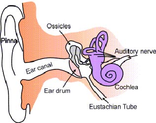
Diagram of Human Ear
The external ear is the fleshy part that helps to collect sounds and funnel them into the auditory canal. The auditory canal extends into the bones of the head but stops at the eardrum, or tympanum. The eardrum is the beginning of the middle ear. Sound waves strike the eardrum and cause it to vibrate. Three small bones behind the eardrum—the hammer, anvil, and stirrup—transfer the vibration to a fluid-filled chamber within the inner ear. This chamber, which is shaped like a tightly coiled shell, is the cochlea. When the fluid vibrates, tiny hair cells in the cochlea are pushed back and forth, providing stimulation that is turned into nerve impulses. These impulses travel on the auditory nerve to the brain.
The ears also contain structures for detecting stimuli that make us aware of our movements and allow us to maintain balance. These structures are called semicircular canals.
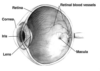
Diagram of Eye
No stimulus provides as much information about the environment as light. Vision depends on the eyes.
Each eye is composed of three layers. The outer layer consists of the sclera and the cornea. The sclera consists of tough, white connective tissue. It helps maintain the eye’s shape and provides a place of attachment for the eye muscles. In the front of the eye, the sclera forms a transparent layer, the cornea. Light will first pass through the cornea, which will focus the light onto the back of the eye. The middle layer of the eye consists of the choroid, ciliary body, and the iris. The choroid contains the blood vessels and becomes the disk-like iris. The iris gives the eye its color and controls the amount of light entering the eye. In the middle of the iris is a small opening, the pupil, through which light will enter the eye. Just behind the iris is the lens. Small muscles attached to the lens cause it to bend slightly. This bending enables the eye to focus on close and distant objects.
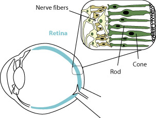
Diagram of Rods and Cones
The inner layer of the eye consists of the retina. The retina is the light-sensing portion of the eye. Special photoreceptors are arranged in a layer in the retina. Rods and cones generate nerve impulses, which travel up the optic nerve to the brain. Rods are extremely sensitive to light and can detect various shades of gray even in dim light. Cones detect color and are sensitive to edges, so they produce sharp images. They produce poorly defined images.
In humans, the field of vision of the two eyes overlaps; each eye sees about one-third of what the other sees. In addition, the two eyes view an object from different angles.
The outer layer of the eye consists of the
The correct choice is D, the sclera and cornea. Choice A is the middle layer and Choice C is the inner layer. Choice D is part of the ear.
The sense of smell depends on receptors that detect specific chemicals in the environment. In the nose, the receptor cells are located in the upper part of the nasal cavity. These receptors contain cilia that extend into the air passageways of the nose and react to chemicals in the air. When they react to chemicals, the receptors are stimulated. This causes impulses to travel up the olfactory nerve to the brain.
The sense of taste is also a chemical sense. The organs that detect taste are the taste buds. About 10,000 taste buds are embedded within the surface of the tongue. Taste buds can also be located on the roof of the mouth and on the lips and throat. The taste buds detect four types of chemicals: sugars (sweet), acids (sour), alkaloids (bitter), and metal ions (salty). Generally, the tip of the tongue is sensitive to sweets, while the back of the tongue is sensitive to bitter tastes. The sides of the tongue are sensitive to sour and salty. The taste buds are stimulated when a chemical dissolved in saliva binds to small hairs that protrude from the tip of the taste bud. This generates an impulse that travels to the brain.
The sense of touch is not found in one particular place. All regions of the skin are sensitive to touch. There are several types of sensory receptors that lie just below the surface of the skin.
Sensory receptors for heat and cold are usually scattered. Those for touch are more concentrated in some parts of the body than others. Pain receptors are located throughout the skin.
The largest sense organ is the
The correct choice is C. The skin has sensory receptors all over it. Choices A, B, and D are sense organs, but none of them are the largest.