The birth of new individuals of a species passes genetic material from generation to generation as a result of sexual reproduction. The genetic material comes from the mother and the father, each providing 50 percent. For this to happen, the gametes must contain half of the needed genetic material. When the sperm and egg come together, the genetic material will combine to make 100 percent.
For this combining of genetic materials to take place, the cells must go through a process called meiosis, which results in the cell containing only half of the needed genetic material. Meiosis only occurs in sex cells.
Meiosis produces haploid gametes. There are two nuclear divisions: meiosis I and meiosis II. In meiosis I, homologous chromosomes pair up, and crossing over occurs. The result is two genetically identical daughter cells. In meiosis II, these daughter cells divide to form four haploid cells. Recall that the gametes have a haploid (n) chromosome number, and the somatic (or body) cells have diploid (2n) number of chromosomes.
In most species there are male and female members. These two genders have reproductive organs that are compatible with each other in order to make reproductions of themselves. In addition to the organs that are needed in reproduction, there are also many hormones that play a large part in the development of the sex organs, producing and releasing eggs and sperm, as well as maintaining a pregnancy.
While the organs used in reproduction are different in males and females, they have complementary components. Essentially the organs consist of:
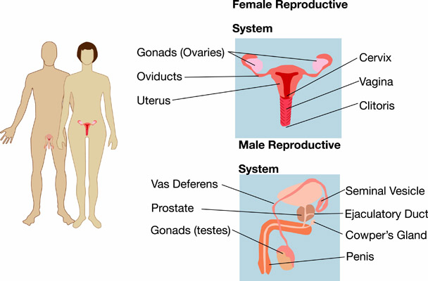
The male reproductive system includes: testes, ductus epididymis, ductus (vas) deferens, ejaculatory duct, urethra, seminal vesicles, prostate gland, bulbourethral glands, and penis.
The scrotum sack, which consists of loose skin and superficial fascia, supports the testes. The testes are paired oval glands that contain seminiferous tubules (where sperm is made), sertoli cells (nourish cells), and Leydig cells (produce testosterone). The testes descend into the scrotum through inguinal canals during the seventh month of fetal development. The temperature of the testes is regulated by the contraction of the cremaster and dartos muscles. These muscles bring the testes up or down as needed into the pelvic cavity.
Spermatogenesis is when immature spermatogonia develop into mature sperm. Mature sperm consist of a head, a midpiece, and a tail. Their function is to fertilize secondary oocytes. During puberty, GnRH (gonadotropin-releasing hormone) stimulates secretion of FSH (follicle stimulating hormone) and LH (leutenizing hormone). LH stimulates production of testosterone. FSH and testosterone stimulate spermatogenesis.
Testosterone’s role is to control growth and maintenance of sex organs, bone growth, protein anabolism, and sperm maturation. It also stimulates the development of secondary sex characteristics, such as facial hair and deepening of the voice.
The duct system consists of seminiferous tubules, straight tubules, and rete testes. Sperm flow out of the testes through efferent ducts. There is also the ductus epididymis, which is a place on the superior portion of the testicle in which the sperm mature. After the sperm mature, they enter the vas deferens, which stores sperm and propels it towards the urethra during ejaculation. Sperm then enter the ejaculatory duct, which is the union of ducts from seminal vesicle and ductus deferens. This acts as a passageway for sperm and secretions of seminal vesicles into the prostatic urethra. There are three parts to the urethra: prostatic, membranous, and spongy (penile).
Accessory sex glands include:
Semen is actually a mixture of sperm and seminal fluid. It provides transportation and nutrients for the sperm, and it helps to neutralize the acidity of the vagina and male urethra (remember, urine also passes through the male urethra).
The main organs of the female reproductive system include: ovaries, uterine (Fallopian) tubes, uterus, vagina and vulva, and mammary glands.
The ovaries are located in the superior part of the pelvic cavity and are lateral to the uterus. Their job is to produce and discharge secondary oocytes (ovulation—the release of an egg for fertilization), and to secrete estrogens, progesterone, relaxin, and inhibin.
An egg is produced through oogenesis, which is the production of haploid secondary oocytes. This process begins in the ovaries. Meiosis II is completed after a secondary oocyte is fertilized by a sperm cell.
The uterine tubes then transport the secondary oocyte from the ovaries to the uterus. The site of fertilization is in the fallopian or uterine tubes. Ciliated cells and peristaltic contractions in the fallopian tubes move the oocyte, or if fertilized, the ovum, to the uterus.
The uterus is the size and shape of an inverted pear and has functions in menstruation, implantation of fertilized ovum, and the development of the fetus. It also acts as a pathway for the sperm to reach uterine tubes. The uterus is held in position by ligaments. There are three main layers to the uterus: outer perimetrium (serosa), middle myometrium, and inner endometrium, the latter which is shed during menstruation.
The vagina acts as a passageway for menstrual flow and sperm, and serves as the birth canal. External genitalia (vulva) include the mons pubis, labia majora and minora, clitoris, vestibule, vaginal and urethral orifices, hymen, and various glands. The perineum is the diamond-shaped area at the inferior portion of the trunk medial to thighs and buttocks.
Females also have mammary glands, which are modified sweat glands that synthesize, secrete, and eject milk. They depend on estrogen and progesterone to produce milk. Milk production is stimulated by the hormones prolactin, estrogen, and progesterone. Milk ejaculation is stimulated by oxytocin.
The monthly menstrual cycle prepares the endometrium of the uterus to receive a fertilized egg. These cycles are controlled by GnRH (gonadotropin-releasing hormone), which stimulates the release of FSH and LH. FSH (follicle-stimulating hormone) stimulates development of secondary follicles and initiates secretion of estrogens. LH (leutenizing hormone) further stimulates the development of follicles, secretion of estrogens, ovulation, formation of the corpus luteum, and the secretion of estrogen and progesterone by the corpus luteum.
Estrogen stimulates the growth, development, and maintenance of secondary sex characteristics, such as additional body hair and breast development, and stimulates protein synthesis. Progesterone works with estrogen to prepare the endometrium to form and the mammary glands for milk synthesis.
During the preovulatory phase, a group of follicles in an ovary undergoes final maturation. One follicle outgrows the others and becomes dominant and the others degenerate. At the same time, endometrial repair occurs in the uterus. Estrogen is the main hormone now.
Around the middle of the menstrual cycle, ovulation occurs and there is the rupture of a mature (Graffian) follicle, which causes the release of a secondary oocyte into the pelvic cavity. There is a surge in LH and there can be some specific signs and symptoms of ovulation, which includes temperature spike and vaginal discharge. Some females feel pain.
During the postovulatory phase, the corpus luteum secretes large quantities of progesterone and estrogen. The endometrium readies for implantation. If there is no fertilization and implantation, the corpus luteum degenerates and hormone levels drop. In particular, there are low levels of progesterone and a discharge of the endometrium. The stratum functionalis of the endometrium is shed, discharging fluid, tissue, mucus, and epithelial cells.
If fertilization does occur, the corpus luteum is maintained by placental hCG and these secrete progesterone and estrogen to support pregnancy and breast development for milk production.
Obviously, changes occur to a female during pregnancy. During the first three months, the corpus luteum produces hCG, progesterone, and estrogen needed to maintain the pregnancy. During the third month, the placenta comes into action and secretes hCG into the blood, causing the release of estrogen and progesterone.
During the pregnancy, the uterus grows with the fetus. In addition, the female increases her blood volume by up to 50 percent. The heart works harder and breathing can become more difficult. There is also weight gain from the fetus, the amniotic fluid, and overall water retention. The breasts also increase in size in preparation for producing milk. The gastrointestinal tract has less motility due to various hormones. Crowding of the bladder, along with the hormones produced in the first trimester cause the need for more frequent urination. There may be changes in the skin, as well.
During parturition (childbirth), there are many hormones interacting to induce the event. For contractions to occur, progesterone levels must be low and estrogen, relaxin, and oxytocin levels must be high. Oxytocin stimulates contractions and relaxin helps in the loosening of the pubic symphysis and dilation of the cervix. Parturition causes a positive feedback loop of hormones until the baby is born.
There are 3 stages of childbirth:
This delivery process is very stressful on both mom and baby. To help counteract the stress, both produce high levels of adrenal medullary hormones like epinephrine. This hormone eases the stress and helps the baby to take its first breath.
During breastfeeding the female’s body produces oxytocin that causes the uterus to contract and return to original size.
Puberty is when secondary sex characteristics begin to develop and the potential for sexual reproduction is reached. In males, there is an increase in LH, FSH, and testosterone levels, resulting in additional body hair and vocal changes. In females, there is an increase in LH, FSH, and estrogen levels, resulting in additional body hair and development of breasts and hips.
As males age, testosterone levels and muscle strength decrease and there are fewer viable sperm. As females age, progesterone and estrogen levels decrease eventually ending with menopause.