In this lesson, you will review the structures and functions of the musculoskeletal system.


There are some skeletal differences between the human genders. Males have longer, thicker limbs and digits; females have narrower ribs, smaller teeth, and less pronounced skull and mandible features. The most notable difference is the pelvis: females have a larger pelvis, which is crucial for reproductive success.
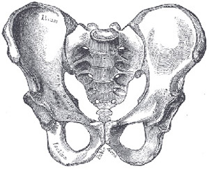
Male pelvis
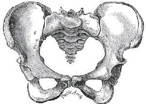
Female pelvis
What type of skeletal system do the majority of vertebrates have?
D is the correct answer. Exoskeletons are exterior and are characteristic of invertebrates, such as arthropods and shellfish. Hydrostatic skeletons operate by pressure and are characteristic of soft-bodied invertebrates, such as corals and leeches. Cartilage is a dense connective tissue and is characteristic of some species of fish, such as sharks and rays.
Cartilage is a dense connective tissue composed of cells called chondrocytes. These cells are dispersed in a firm, gel-like, ground substance called the matrix. There are no blood vessels in cartilage; therefore nutrients are diffused through the matrix.
Cartilage is found in the nose, the ears, the throat, the rib cage, between intervertebral disks, and in the joints.
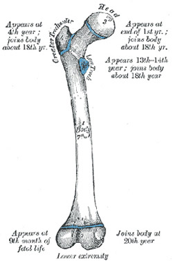
Many vertebrates have hard endoskeletal connective tissue we call bone. Collectively, all the body’s bones comprise the skeleton. The longest bone is the femur and the smallest is the stapes in the middle ear.
Bone is derived from the mesoderm during embryonic development. Bones function to support the body’s structure, protect internal organs, and facilitate movement in conjunction with muscles. Bones are also involved in cell formation, calcium metabolism, and mineral storage.
There are two types of bones in the vertebrate body plan, the axial skeleton and the appendicular skeleton. The axial skeleton consists of bones along the axis, the head, vertebrae, and ribs. The appendicular skeleton, on the other hand, consists of everything else: clavicles, scapulae, pelvis, and the upper and lower limbs.
Ligaments are short bands of tough, fibrous connective tissue. Their composition is long, stringy collagen fibers that are slightly elastic. Ligaments connect one bone to another, forming joints. Ligaments provide mobility, limit mobility, and prevent mobility. Because of their slight elasticity, ligaments gradually lengthen under tension. Dislocated joints need to be set quickly, as lengthening from the dislocation results in a compromised joint allowing for further dislocations. Double-jointed individuals have very elastic ligaments that allow a certain amount of contortion.
Similar to ligaments, tendons are tough bands of fibrous connective tissue connecting muscle to bone. A tendon originates where it joins a muscle. The muscles’ collagen fibers are continuous with the tendon’s collagen fibers, which insert into a bone and integrate into the bone tissue.
Which structure diffuses nutrients through cartilage?
The correct answer is D. Cartilage has no blood vessels; therefore choices A and B are incorrect. Lymph vessels transport lymph and are associated with the immune system; therefore, choice C is incorrect.
Which structure connects bone to bone?
C is the correct answer. Choices A and D are incorrect because tendons connect muscle to bone. Choice B is incorrect because cartilage is a dense connective tissue that is connected to bone, but does not connect bone to bone.
Muscle is contractile tissue. The contraction of many muscles at the right time is the only way locomotion is possible. Muscle contraction occurs with the shortening of a muscle fiber (cell).
Muscle is also involved in moving substances throughout the body. Conscious effort of the organism stimulates most muscle contractions. A signal is relayed from the brain in the form of an action potential, which travels through the nervous system and arrives at the motor neuron that innervates (supplies with nerves) the muscle fiber. However, unexpected stimuli can cause a muscle reflex resulting from a signal from the spinal cord. Such reflexes are fast and unconscious. Autonomic muscle contractions are also unconscious and are what keeps the heart beating.
There are three types of muscle contractions: heart, smooth muscle, and skeletal muscle. The following steps explain skeletal muscle contractions.
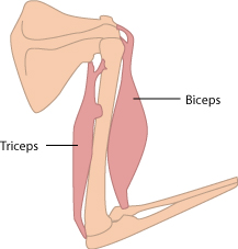
Muscles only have the ability to contract, meaning they can pull but they cannot push. This is the reason muscles are found in opposing pairs, or antagonistic pairs. So when a muscle contracts to bend an arm or knee, there is an opposing muscle ready to contract and straighten the arm or knee.
An example of an antagonistic pair of muscles is the biceps and triceps muscles in the arms. The arm bends when the biceps muscle contracts; it straightens when the triceps muscle contracts.
Another example of an antagonistic pair of muscles is the circular and radial muscles in the eye. The iris controls the size of the pupil for light to enter the eye. When the circular muscle of the pupil narrows, less light enters; when the radial muscle widens, more light enters.
What is the term used for muscles in opposing pairs?
C is the correct answer. Choices A, B, and D are synonyms for the word “opposing”.