 username@email.com
In this lesson, you will review the structures and functions of animal cells.
Despite their microscopic size, cells contain numerous internal structures called organelles. The structure and function of organelles is often highly complex, and nearly all organelles are involved in intricate biochemical reactions that maintain the living cell. The major organelles in animal cells are outlined below.

A diagram of a cross-section of a mitochondrion, with labels

A diagram of organelles involved in protein sythesis and transport
Cytoplasm is the living contents inside a cell, excluding the nucleus and the large vesicles. The cytoplasm contains a clear, colorless liquid termed the hydroplasm in which the organelles occur. Cytoplasm is about 90 percent water, but that water is a rich solution of ions, especially potassium, sodium, and chloride. Of course, the cytoplasm is bounded and contained within the plasma membrane.
A cell’s cytoskeleton helps support the cell and keep it in shape. The cytoskeleton consists of interconnected protein filaments in the cytoplasm. The cytoskeleton not only maintains the cell’s three-dimensional shape, it also anchors its organelles and helps the cell move.
Three basic types of structures make up the cytoskeleton:
Which of the following is a cytoskeleton structure?
The correct answer is A. The other three choices are organelles.
The plasma membrane encloses and protects the contents of the cell. As with all living things, however, cells do interact with their environment. This means that material must be able to pass into and out of the cell through the plasma membrane. The thin, seemingly simple plasma membrane is actually a formidable, complex structure that serves as an effective barrier that keeps unwanted material out of the cell while permitting the entry of useful substances. This makes the plasma membrane selectively permeable—only allowing in needed substances and keeping out unwanted material.

The plasma membrane selectively allows transport of substances into and out of the cell. Intrinsic proteins on the membrane enable passage of needed large, water-soluble molecules and ions across the plasma membrane and into the cell. Some of these proteins produce channels, like tiny pores, through which molecules can pass. Some channels are akin to gatekeepers that open only when a particular chemical signals its “intent” to enter the cell. Gap junctions are protein channels that permit communication between cells.
Other intrinsic proteins simulate ferries that carry a particular molecule across the plasma membrane. Some intrinsic proteins act on the molecule chemically to make it appear more “friendly” to the membrane. Still other proteins pump in the needed molecules.

Most molecules pass through the plasma membrane via diffusion, in which a molecule or substance moves, without any input of energy, from a region where it occurs in high concentration to a region where it occurs in low concentration. Molecules move via diffusion until the entire region reaches equilibrium in terms of the concentration of the particular molecule. (The spread of an aroma through a room is an example of diffusion). The concentrations for most substances inside and outside the cell differ and materials diffuse through channels via passive transport until equilibrium is reached. In some cases, intrinsic proteins assist in moving molecules along the concentration gradient; this process is called facilitated diffusion.
There are times, however, when a molecule must be moved into or out of a cell against the concentration gradient; that is, it must be moved from a region of low concentration or equilibrium to a region of high concentration. An input of energy is needed to accomplish this. Membrane proteins provide this energy boost to move molecules via the process of active transport.
The sodium-potassium pump is a typical active transport system in cells. In most cells, sodium is maintained in low concentrations inside the cell, though its concentration is higher outside the cell. The reverse is true of potassium. ATP (adenosine triphosphate) fuels the active transport of these ions across the plasma membrane to maintain the requisite cell concentrations. Both sodium and potassium are carried by a transport protein, which occurs in two forms—one for each ion. The pump acts as follows: A sodium ion binds to form 1 of the protein while ATP attaches a phosphate group. This causes the protein to change into form 2 and, while shape-shifting, to deposit the sodium ion outside the plasma membrane. The protein now grabs a potassium ion and releases its phosphate group, which transfigures the protein back to form 1. The protein then releases the potassium ion inside the cell.
Diffusion, facilitated diffusion, and active transport are used to move substances across the membranes of organelles, as well as across the plasma membrane.

A diagram of Na-K pump
What triggers the transfiguration of the intrinsic protein involved in the sodium-potassium pump?
The correct choice is C. The addition or removal of a phosphate group triggers the intrinsic protein to change shape, and causes it to pick up or deposit either sodium or potassium in or out of the cell. A sodium ion binds to an intrinsic protein that is already primed to receive it; it does not trigger changes in the intrinsic protein, so choice A is incorrect. The same holds true for the binding of potassium to the intrinsic protein, therefore choice B is incorrect. Intrinsic proteins are involved in active transport, which is transport against ion concentration on either side of the cell membrane. Thus, the ion concentration does not trigger changes in the intrinsic protein that enable it to carry ions against the ion gradient, so choice D is incorrect.
Proteins and amino acids are large particles, and they’re too big to cross the plasma membrane via diffusion or by active transport. Yet cells need these substances. What to do? Cells use vacuoles, or vesicles, to get these necessary particles inside.
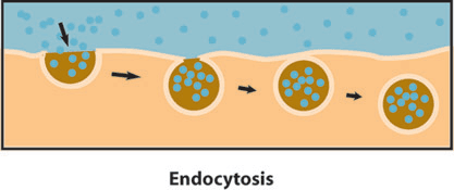
Endocytosis is a transport process that carries large particles into the cell. In endocytosis, a protein particle, for example, attaches itself to a special area on the outside of the plasma membrane. Attachment triggers the membrane to bulge inward to form a kind of pouch, or vesicle. The pouch enlarges until the particle is completely enclosed in it. Then the vacuole and its contents are released into the cytoplasm.


During facilitated transport, intrinsic proteins
C is the correct choice. In facilitated transport, or facilitated diffusion, intrinsic proteins help move molecules in the direction of the concentration gradient. Carrying ions in a direction against the concentration gradient, which would be from a region of high concentration to low concentration, is active transport, not facilitated transport, therefore choice A is not correct. Choice B is incorrect because it is gap junctions that permit chemical communication among cells. Proteins are too large to move through the plasma membrane via diffusion, even facilitated diffusion. They must be moved via active transport, so choice D is incorrect.
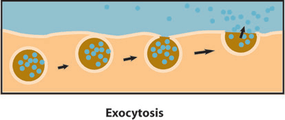
In exocytosis, transport vesicles are created in
The correct choice is A. Exocytosis creates vesicles in the Golgi bodies, which encase the particle in a membrane that attaches to the plasma membrane, from where the particle is expelled. The SER is involved in the synthesis of cholesterol, not in exocytosis, so choice B is incorrect. Intrinsic proteins assist in the transport of needed molecules into the cell. Though in active transport, intrinsic proteins help rid the cell of sodium ions, this is not exocytosis, so choice C is incorrect. ATP is the energy source of the cell, created in the mitochondria. Although ATP fuels all cell functions, it is not directly involved in exocytosis, therefore choice D is incorrect.
Endocytosis is triggered by
B is the correct choice. When a needed particle attaches to a receptor on the cell surface, endocytosis is triggered to allow the particle into the cell. ATP remains inside a cell and is not activated chemically by particles needed inside the cell, therefore choice A is not correct. Choice C is incorrect because the endoplasmic reticulum synthesizes necessary materials for the cell. It is not involved in transport into the cell. Endocytosis involves the entry of needed particles, such as proteins, into the cell. Choice D is incorrect because it describes exocytosis.
You have read that cells get their energy from ATP (adenosine triphosphate). But what is ATP and where does it come from? ATP is the molecule of choice for energy transfer in all cells. ATP stores the energy that is used in cellular processes in the high-energy chemical bonds between its three phosphates. The breakdown of ATP breaks the phosphate bonds, releasing energy and making it available to the cell. The formula for this reaction, which works in reverse when phosphate is added to ADP to make ATP, is
ATP + H2O –> ADP + Pi + energy
Often, the breakdown of ATP does not release inorganic phosphate, but instead transfers it, with the aid of an enzyme, to another molecule. This process is called phosphorylation. Thus, the phosphorylation of ADP creates ATP. Getting ATP from glucose is a multi-step process. The first step is glycolysis, which breaks down glucose; the second step is respiration, which itself consists of two steps: the Krebs cycle and electron transport chain. Both oxidation (the loss of an electron) and reduction (the addition of an electron) may be used to create energy from glucose.
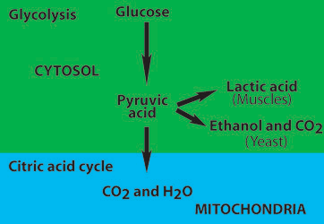
Glycolysis, which occurs in the cell cytoplasm, is an anaerobic process that entails the splitting, or lysing, of glucose. There is a specific sequence of nine chemical reactions in glycolysis, each catalyzed by a particular enzyme. The overall purpose of glycolysis is to break down the carbon bonds in glucose and to use the released energy to produce fuel, or energy, for the cell (in the form of ATP):
glucose + oxygen –> carbon dioxide + water + energy
C6H12O6 + 6 O2 –> 6 CO2 + 6 H2O + energy
In glycolysis, a phosphate group is transferred from an ATP molecule to a glucose molecule. The glucose molecule then splits apart, and energy is produced as NAD (nicotinamide adenine dinucleotide—a hydrogen carrier). NAD is reduced to NADH (hydrogen having been obtained from PGAL, phosphoglyceraldehyde). Finally, molecules of ADP are phosphorylated to become ATP.

A full schematic of Glycolysis
Glycolysis yields 2 ATP plus 2 molecules of pyruvic acid. ATP do not move easily across the inner mitochondrial membrane, so its electrons must be carried across. Once this occurs, more ATP molecules are produced. In total, glycolysis yields 8 ATP.
Cellular respiration is the oxidation of food (glucose) by cells. Cellular respiration entails the further breakdown of glucose to fuel cell function. In cellular respiration, a sequence of reactions oxidizes pyruvic acid, which is produced in the later steps of glycolysis, to yield energy, carbon dioxide, and water. Cellular respiration occurs in the mitochondria and involves two steps: the Krebs cycle and electron transport chain.
The prelude to the Krebs cycle is often called the transition reaction. The first step in cellular respiration entails the oxidation of pyruvic acid. The carbon is removed from the three-carbon pyruvic acid and forms 2 CO2. Two two-carbon acetyl groups are left (2 CH3CO). The pyruvic acid’s hydrogen atoms are transferred to hydrogen-carrying molecules of NAD to form 4 NADH. Each acetyl group bonds with coenzyme A (a compound made up of nucleotides and forms of vitamin B) to form the substance acetyl coenzyme A, the key compound that links glycolysis with the Krebs cycle. Converting pyruvic acid to coenzyme A yields 6 ATP.

A schematic of acetyl coenzyme A production
In the Krebs cycle, carbons from the acetyl group (in acetyl coenzyme A) are oxidized to create carbon dioxide, and hydrogen atoms are transferred in an electron transport process. Coenzyme A is involved both in the oxidation of pyruvic acid and in the Krebs cycle.
When the two-carbon acetyl group enters the Krebs cycle, it is combined with a four-carbon compound (oxaloacetic acid) to form the six-carbon compound citric acid. During this process, two of the six carbons are oxidized to carbon dioxide. Some of the energy released during oxidation and breaking of the carbon-carbon and carbon-hydrogen bonds is used to change ADP to ATP, and some of the energy is used to transform NAD to NADH. Remaining energy is used to reduce another electron carrier, FAD (flavin dinucleotide) into FADH2. Oxygen is not used during the Krebs cycle. All the electrons and protons released are picked up by the NAD+ and FAD. The total number of ATP molecules produced in the Krebs cycle is 24 per molecule of glucose.
The formula for Krebs cycle is:
oxaloacetic acid + acetyl Coenzyme A + ADP + P + 3NAD + FAD ⇒ oxaloacetic acid + 2CO2 + Coenzyme A + ATP + 3NADH + FADH + 3H+ + H2O


The main carriers in the electron transport chain are known as cytochromes, which are made up of protein and a porphyrin ring. At each step, a different cytochrome, designed specifically to carry an electron at a particular energy level, carries the electron to the next lower level, ending finally with low-energy oxygen. When, at the end of the downward slide, the electrons link up with oxygen, it then combines with protons (hydrogen ions) to yield water.
At each lower step in the chain, the energy released by a pair of electrons is sufficient to transform, or phosphorylate one ADP to one ATP molecule. Once created, an ATP molecule is moved across the mitochondrial membranes. Simultaneously, an ADP molecule moves into the mitochondrion to begin its transformation by phosphorylation into ATP. You can see that the transformation of ADP into ATP, and vice versa, is a perpetual cycle in which a cell creates and uses the fuel that keeps it going.
In summary, glycolysis and cellular respiration yield 38 molecules of ATP to energize the cell.