In this lesson, you will review the discovery of DNA and the mechanics of molecular biology.
Gregor Mendel was an Austrian monk who brought careful, systematic observation to the study of genetics, which is the study of how characteristics are passed form parent to offspring. Mendel first proposed a particulate or gene model for inheritance. According to his model, parents pass on distinct inheritable units, called genes, to their offspring. This is the basis of the modern science of genetics.
Mendel formulated four laws of genetics.
For example, a white flower is crossed with a red flower. They produce offspring that are only white flowers. These white flowers are then crossed. Their offspring include both red and white flowers. What is the simplest explanation for this distribution of color variation?
Assume there are two alleles for the flower color gene; call them B and b. B shows incomplete dominance. That is, homozygous BB would produce white flowers, heterozygous Bb would produce white flowers, and homozygous bb would produce red flowers. Therefore, both parents must have the BB genotype (white) and the bb genotype (red) to produce only white flowers. By crossing the resulting white flowers to produce both red and white indicates that the offspring were heterozygous, Bb. When crossed, the Bb genotype is capable of producing both BB (white) and Bb (red) offspring.
For decades following Mendel’s discoveries, biologists had thought about the details of the mechanism by which genetic information was transmitted. Biologists surmised that chromosomes, which are visible using an ordinary light microscope during cell division, played some role in the transmission of genetic information. During cell division, the chromosomes appear to aggregate, then separate into pairs. Around 1902, Walter Sutton, Theodore Boveri, and others had proposed a chromosome theory of inheritance. According to this theory, Mendelian genes are located at specific spots on chromosomes, which are duplicated during cell division.
Thomas Hunt Morgan was the first to identify a particular Mendelian gene with a specific chromosome. Just as Mendel studied pea plants because they had simple, easily observable characters, Morgan studied a particular species of fruit fly, Drosophila melagonaster, that has a number of easily observable characters. These flies are ideal laboratory subjects because they breed prolifically and are generally harmless if they escape the lab. A single breeding pair of flies can have hundreds of offspring, producing a new generation every two weeks.
After many generations, Morgan noticed a single male fly with white eyes instead of the normal red. This character is now known to be the product of a mutation in the gene for eye color. Morgan mated the white-eyed fly with a red-eyed female. The F 1 offspring all had red eyes. When this generation was mated, the F 2 generation showed the typical 3:1 phenotypic ratio predicted by the Mendelian model. There was one startling difference, however. The white-eyed flies were all males. Somehow, reasoned Morgan, a fly’s eye color was linked to its sex. He found that the white-eye gene is located on the X chromosome; therefore, males only have one copy of the gene for eye color.
Subsequent discoveries have shown that there are many linked genes. These are genes that are carried on the same chromosome, so they are typically inherited together; the chromosome is passed along as a unit. There are exceptions to this pattern, since linked genes sometimes appear to be inherited separately. Morgan theorized that some mechanism existed that allowed the exchange of genetic information between chromosomes.
Color-blindness is a sex-linked (x) recessive trait and male dominant. A man with color blindness marries a woman with normal vision; however, the woman’s father also has color blindness. If the couple has a daughter, what is the probability that she carries the recessive trait?
C is the correct choice. The woman, with two X chromosomes, inherited the recessive, sex-linked trait from her father’s X chromosome. Her husband, with an X and a Y chromosome, has the disorder, so his X chromosome also contains the recessive allele. Constructing a Punnett square will show that the probability of any children having blindness is 50%, or 0.50, regardless of sex. If the Punnett square is constructed properly, the other choices are rendered incorrect.
Many studies had narrowed down the possible composition of the genetic material to a few possibilities: nucleic acid and various proteins. In 1868, the Swiss biologist Friedrich Miescher began the first careful, systematic studies on the nuclei of cells. Using cells collected from discarded surgical bandages, Miescher detected a substance that he named nuclein, which is a mixture of DNA and RNA.
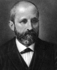
In 1869, Miescher isolated pure DNA from the nuclei of salmon sperm cells. In 1889, Miescher’s pupil, Richard Altmann, named the substance nucleic acid. Biologists quickly identified nucleic acid as a long-chain polymer. They also realized that nucleic acid was a mixture of two different polymers—one containing ribose and the other deoxyribose. DNA was found to exist only in the chromosomes of cells, but it did not seem to have the molecular complexity necessary to be the carrier of genetic information. The proteins found in cell nuclei, on the other hand, appeared to have the necessary variety of forms and complexity.
Since proteins had great heterogeneity and specific functions, they remained the best candidate for the carrier of genetic information until the 1940s. Little was known about the molecular structure of nucleic acids. The first hint that DNA was responsible came from Frederick Griffith. He killed a virulent form of Streptococcus pnuemoniae with heat, and mixed the remains with a harmless form of the same bacteria. Some of the harmless bacteria became pathogenic. Furthermore, the descendents of the disease-causing bacteria were also pathogenic. Apparently, new genetic information had been introduced into the nonpathogenic bacteria.
Griffith’s experiment launched a 14-year effort to find the substance responsible for this transfer of genetic information. In 1943, Oswald Avery purified various chemicals from the killed pathogenic bacteria and tried mixing each in turn with the live nonpathogenic strain. Only DNA worked. However, this idea met with resistance because proteins, due to their complexity, were still considered the best candidate. No one could figure out how DNA, thought to be a simple polymer, could carry genetic information.
In subsequent years, evidence from experiments with viruses, which contain only DNA or RNA, strongly suggested that viral DNA was capable of reprogramming normal cells. A hint of how DNA might carry genetic information was found by Erwin Chargaff. He discovered that the base sequences found in DNA molecules (adenine, A; thymine, T; guanine, G; cytosine, C) differed among different organisms. Chargaff also found some peculiar regularity in the ratios of different bases. In humans, the percentage of A and T are approximately the same (around 30%) and the percentage of G and C are approximately the same, around 20%. These A = T and G = C regularities, known as Chargaff’s rules remained unexplained until the discovery of the structure of DNA.
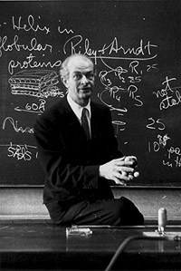
Once DNA was identified as the genetic material, the race was on to discover its structure. There are five names generally associated with the discovery of the structure of DNA and there is considerable debate as to who should receive full credit for the discovery. Like all science, the truth is usually somewhere in the middle and whoever receives credit for the discovery ultimately based his or her work on the discoveries of many others. The five people instrumental to the discovery of DNA are: Linus Pauling, Maurice Wilkins, Rosalind Franklin, James Watson, and Francis Crick.
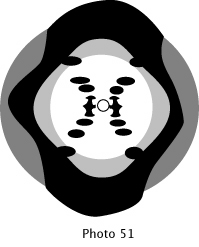
Pauling clearly was the first to propose a helical structure, but had a three-strand helix with the bases pointed outward. Pauling had asked to see the new, sharper, x-ray crystallographic photographs taken by Wilkins and Franklin. The most famous of the photographs taken by Wilkins and Franklin is known as Photo 51. However, Pauling’s request was denied and he had to work with older published photographs that were too blurry to show details. Furthermore, Pauling was prohibited from attending a London conference that might have provided him with revealing information.
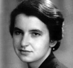
Franklin, Crick, and Watson were colleagues working on determining the structure of DNA. However, egos and personalities have been the speculation for why Franklin did not share credit for the discovery. In addition, Franklin was deceased when the Nobel Prize was awarded for the DNA structure discovery, and the prestigious prize is not awarded posthumously.
Thus, Watson, Crick, and Wilkins shared the Nobel Prize for determining the structure of DNA.
Which generally unrecognized scientist did research that assisted Watson and Crick’s discovery?
The correct choice is C. Wilkins shared the Nobel Prize with Watson and Crick. Pauling conducted DNA research, but encountered obstacles that prevented him from further success. Mendel’s work preceded Watson and Crick’s research, and Mendel is recognized as the Father of Genetics.
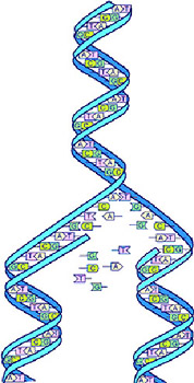
Watson and Crick constructed an actual physical model of DNA. Today, the molecule is most often represented as a computer graphic.
DNA consists of pairs of molecules, which wrap around each other. Imagine a rope ladder that has rigid rungs. The ropes represent the sugar-phosphate backbones and the rungs represent nitrogenous base pairs. Franklin’s x-ray data indicated that the molecule makes one twist every 3.4 nm, and the bases are stacked just 0.34 nm apart. A rope ladder twisted so that there are 10 rungs per twist provides the basic model for DNA.
Watson and Crick used wire to construct scale models that would match the x-ray data. They first tried pointing the bases outward, but they could not get this model to work. So they swiveled the bases inside the spiral. This made sense, because the bases are somewhat hydrophobic and putting them inside the spiral moved them away from water molecules.
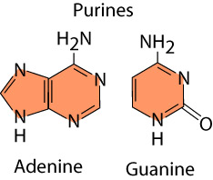
Crick and Watson also assumed the bases would pair with each other. But this also did not fit the x-ray data, because the data showed that the helix had a uniform diameter. Adenine and guanine are purines, nitrogenous bases with two organic rings.
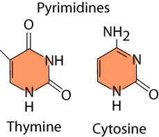
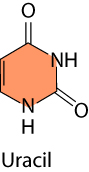
In contrast, thymine and cytosine are pyrimidines, nitrogenous bases with a single ring. There is one other nitrogenous base, the pyrimidine uracil, which is equivalent to thymine and is in RNA.
Watson and Crick also determined that chemical side groups on each base form hydrogen bonds with its paired base. Adenine and thymine can form two hydrogen bonds, while guanine and cytosine can form three hydrogen bonds. The combination of size and hydrogen bonding means that only A-T bonds and G-C bonds can form. So, in addition to matching the x-ray data, the Watson-Crick model explained Chargaff’s rules.
The DNA molecule is a very long, thin polymer. The molecule itself is so thin that it is just barely visible with an electron microscope. Yet the molecule is very long, compared to its width. A typical nucleotide sequence comprising a gene on a DNA molecule would be only a few centimeters long. The total length of human DNA, if it could be stretched out into a single strand, would be several meters long. This molecule, with its billions of nucleotide bases, has the complexity necessary to be a blueprint for a living organism.