For the next lesson, we will review the next highest level of organization for an individual organism, the organ system. An organ system consists of two or more organs that work together. In most animals, there are 5 to 8 organ systems that are all connected and interrelated. Because we are most familiar with our own systems, the following review will concentrate on vertebrates (mammals specifically, and humans in particular) systems. However, most of these organ systems function similarly in all animals. In this lesson, we will review the structures and functions of the musculoskeletal, circulatory, nervous, digestive, excretory, respiratory, urinary, and reproductive systems.
In the previous lesson, we reviewed the important types of cells found in animals and set the stage for higher orders of organization within the animal body plan.
The musculoskeletal system is an organ system that enables animals to move by using muscles and skeleton in concert. Vertebrate bones also support and protect internal organs, and in many organisms bones also produce blood cells and store fat and minerals.
The human musculoskeletal system consists of the skeleton, made up of bones attached by joints, and skeletal muscle attached to the skeleton by tendons. Many animals have hydrostatic skeletons and move by contracting muscles surrounding a fluid-filled pouch, creating pressure that causes movement. Other animals, most notably the arthropods, have muscles attached to an exoskeleton that coincidentally acts as body armor.
There are three basic kinds of skeletal systems, external (exoskeleton), internal (endoskeleton), and fluid (a hydrostatic skeleton). Because larger external skeletal systems can support less weight than endoskeletons of the same size, many animals like the vertebrates, have evolved internal skeletal systems. Exoskeletons are found in arthropods and many mollusks. An internal skeletal system is made up of bones or cartilage within the body, moved by the muscular system. Some organisms like sharks and rays have a skeleton consisting only of cartilage without calcified bones. Hydrostatic skeletons are similar to a water-filled balloon. Located internally in cnidarians (coral, jellyfish, etc.) and annelids (leeches), among others, these animals can move by contracting the muscles surrounding the fluid-filled pouch, creating pressure within the pouch that causes movement. Animals such as earthworms use their hydrostatic skeletons to change their body shape as they move forward from long and skinny to short and stumpy.
The skeleton functions not only as the support for the body but also in the manufacture of blood cells that takes place in bone marrow. It is also necessary for protection of vital organs and is needed by the muscles for movement.
There are two types of bones in the vertebrate body plan, the axial skeleton and the appendicular skeleton. The axial skeleton consists of bones along the axis, the head, vertebrae, and ribs. The appendicular skeleton, on the other hand, consists of everything else: clavicles, scapulae, pelvis, and the upper and lower limbs. There are also many differences between female and male human skeletons. While men have longer and thicker limbs and digits, women have larger pelvises, narrower ribs, smaller teeth, and less pronounced skull and mandible features. Most notable is the difference in the pelvis, a strongly selected trait that is crucial for women’s reproductive success.
Blood is the body’s internal transportation system. The circulatory system connects the various muscles and organs of the body with one another. It consists of the heart, blood, and blood vessels. The circulatory system has four functions:
The heart is a hollow, muscular organ that contracts at regular intervals, forcing blood through the circulatory system. The walls are made of three layers of tissue. The outer and inner layers are epithelial tissue. The middle layer is cardiac muscle tissue.
The pattern of circulation in the human body includes two separate circulatory loops. The right side of the heart pumps blood from the body into the lungs, where deoxygenated blood gives up carbon dioxide and picks up oxygen. This is the pulmonary circulation loop. The left side of the heart pumps oxygenated blood from the lungs to the rest of the body. This is the systemic circulation loop.
The heart is enclosed in a protective sac of tissue called the pericardium. A septum divides the two sides of the heart, preventing the mixing of the deoxygenated blood with the oxygenated blood.
Oxygenated blood flows from the lungs into the left side of the heart through the pulmonary veins and empties into the heart’s collection chamber, the left atrium. From the left atrium, the blood flows into the left ventricle. When the heart starts to contract, the atrium contracts first. This pushes the remaining blood into the ventricle. Then the ventricle contracts. The blood is prevented from going back into the left atrium by a one-way valve called the mitral valve. Next the blood enters the aorta, which is the largest artery in the body. Once in the aorta, the blood is again prevented from moving backwards by another large valve, the aortic valve. Many arteries branch from the aorta and carry oxygen-rich blood to the body. The first arteries to branch off are the coronary arteries, which carry oxygenated blood to the heart muscles.
After delivering oxygen to the body’s cells, blood returns to the heart through veins. Two large veins collect all the oxygen-poor blood. The superior vena cava drains blood from the upper body, while the inferior vena cava drains blood from the lower body. These veins empty into the right atrium of the heart. The blood then flows into the right ventricle through the tricuspid valve. As the right ventricle contracts, the blood is sent through the pulmonary valve and into the pulmonary arteries. These arteries carry the blood to the lungs. The blood then returns to the left atrium, full of oxygen.
The heart does not contract in a single motion. Instead, the contraction spreads out over the heart like a wave. A smaller cluster of cells, the sinoatrial node, embedded in the right atrium, initiates this wave. These cells act as a pacemaker, setting the pace for the heart rate. The impulse travels to the left and right atria, causing both of them to contract almost simultaneously. The right and left ventricles will also contract simultaneously when the impulse reaches them. A tenth of a second usually passes before the lower part of the heart starts to contract.
These contractions cause the chambers to squeeze the blood, pushing it in the proper direction along its path. In most people at rest, the heart rate is between 60 and 80 beats per minute. The heart rate will increase during exercise in order to pump oxygen-rich blood through the body more quickly.
Arteries carry blood from the heart to the body tissues. The walls of arteries are thicker than that of veins. Except for pulmonary arteries, all arteries carry oxygenated blood. The wall of an artery is made up of three layers of tissue. This allows the artery to expand as blood is pumped into it. Smaller branches of arteries are called arterioles.
Arterioles branch into networks of capillaries, which are tiny blood vessels. The capillaries is where the real work of circulation is done. The walls of capillaries consist of only one layer of cells. This makes it easy for oxygen and nutrients to diffuse from the blood into tissues. Capillaries are extremely narrow — blood cells must move along them in single file.
The flow of blood moves from capillaries into veins. Veins collect blood from the body and carry it back to the heart. Veins are lined with smooth muscle; however, the walls are thinner and less elastic than arteries. The walls of veins are able to stretch out, thus reducing the resistance the flow of blood encounters on its way back to the heart. Large veins contain one-way valves that keep blood from flowing backward. The largest vein in the body is the vena cava, which leads into the heart.
The cardiovascular system is very leaky. Fluids are forced out of the capillaries by the pressure generated when the heart pumps. A network of vessels, known as the lymphatic system collects the fluid and returns it to the circulatory system. The fluid, called lymph, collects in lymph capillaries and flows into lymph vessels. The fluid is returned to the circulatory system at an opening in a vein located under the left clavicle, just below the shoulder.
The smallest blood vessels are
The correct answer is B. Capillaries are so small and narrow, the blood cells have to move through them one-at-a-time. Veins are the next smallest and arteries are the largest. Lymph vessels are not blood vessels.
Blood is a liquid connective tissue that has multiple functions:
The human body contains four to six liters of blood. Fifty-five percent of blood is made up of a fluid portion called plasma. The remaining forty-five percent consists of cells.
Plasma is a yellowish fluid that is 90 percent water and 10 percent dissolved fats, salts, sugars, and proteins. The plasma proteins are divided into three types: albumins, globulins, and fibrinogen. Albumins regulate osmotic pressure. Globulins include antibodies that help fight off infection. Fibrinogen helps the blood to clot. Plasma also carries nutrients, hormones, and waste products.
The cellular portion of the blood includes several types of highly specialized cells and cell fragments. They are red blood cells, white blood cells, and platelets.
Red blood cells, also called erythrocytes, are the most numerous of the blood cells. They are shaped so that they are narrower in the center than along the edges. Erythrocytes are produced from cells in bone marrow that gradually fill with hemoglobin>>. Hemoglobin is the iron-containing protein that carries oxygen from the lungs to the tissues of the body and also gives the erythrocytes their characteristic red color. Red blood cells lack nuclei and organelles and normally stay in circulation for 120 days before they are destroyed by specialized white blood cells in the liver and spleen.
About 90 percent of plasma is composed of
D is the correct choice, because plasma is the liquid portion of blood. The other 10 percent of plasma consists of dissolved fats, salts, sugars, and proteins.
Blood Pressure
Blood moves through the vessels because it is under pressure. This pressure is produced by the contraction of the heart and by the muscles surrounding blood vessels. Blood pressure is a measure of the force that blood exerts against a vessel wall. The low pressure that occurs during relaxation of the heart is diastolic pressure. The higher pressure that results when a pulse of blood is forced into the arterial system is systolic pressure.
What is the function of lymphatic vessels?
B is the correct choice. The lymphatic system collects the fluid that has leaked out of the circulatory system and returns it to the blood. Blood is transported by the circulatory system. The immune system produces antibodies. Blood clotting is controlled by platelets in the circulatory system.
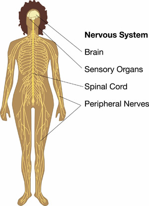
The nervous system controls and coordinates essential functions of the human body. This system receives and relays information about activities within the body, and monitors and responds to internal and external changes.
Cells that carry messages throughout the nervous system are called neurons. These messages are called nerve impulses. Neurons are classified into three types according to the directions in which the impulses move.
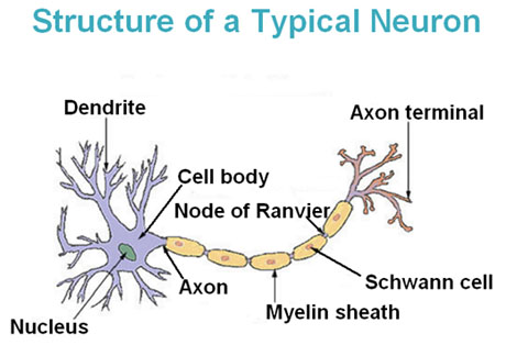
The largest part of the neuron is the cell body. The cell body contains the nucleus and most of the cytoplasm; therefore, most of the cell’s metabolic activities occur here. Spreading out from the cell body are branched extensions called dendrites. They carry impulses toward the cell body. The long fiber that carries impulses away from the cell body is the axon, which ends in a series of swellings called axon terminals.
Bundles of neurons are called nerves. The nerves contain a large number of independent communications channels. They are also composed of many supporting cells that form nervous tissue. Schwann cells wrap around axons forming a fatty, insulating covering called a myelin sheath. There are small gaps, called nodes, between the myelin sheaths. As an impulse moves down an axon, it will jump from node to node. This increases the speed of the impulse.
A nerve impulse is a flow of electrical charges along the cell membrane of a neuron. The strength of an impulse is always the same. The minimum level of a stimulus that is required to activate a neuron is the threshold.
At the axon terminals, the neuron may make contact with dendrites of another neuron, with a receptor or an effector. Receptors are special sensory neurons in sense organs that receive stimuli from the external environment. Effectors are muscles or glands that bring about a coordinated response. The point of contact where an impulse is passed from one cell to another is called a synapse. Tiny vesicles filled with chemicals lie at the synapse. These vesicles are called neurotransmitters, which are used by one neuron to signal another.
Which type of neuron is responsible for transmitting impulses to the central nervous system?
A is the correct choice, because sensory neurons carry signals from sense organs to the brain and spinal cord. Interneurons connect sensory and motor neurons and carry impulses between them. Receptor neurons are special sensory neurons in sense organs that receive stimuli from the external environment. Motor neurons carry impulses from the brain and spinal cord to muscles or glands.
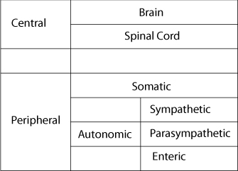
The human nervous system is divided into two major divisions: the central nervous system and the peripheral nervous system. The central nervous system serves as the control center of the body. It consists of the brain and the spinal cord. Its functions are relaying messages, processing information, and comparing and analyzing information. The peripheral nervous system consists of nerves and ganglia, including those of the brain and spinal cord.
The brain contains approximately 35 billion neurons and in order for the brain to function, it must have a constant supply of food and oxygen.
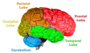
The largest part of the brain is the cerebrum. It is responsible for all of the voluntary activities of the body. It is also the sight of intelligence, learning, and judgment. The cerebrum is divided into two hemispheres, right and left, by a deep groove known as the corpus callosum. The surface of the cerebrum has numerous folds. Each hemisphere is divided into regions called lobes>>, which are named for the skull bones that cover them.
Not surprisingly, the brain is not just uniform gray matter, it has many parts and distinct areas. The right hemisphere is generally associated with creativity, communication, and artistic ability while the left hemisphere is more dedicated to analytical, spatial, and mathematical ability. The cerebrum consists of two surfaces. Much of the activity occurs in the cerebral cortex, which is composed of gray matter. The gray matter contains densely packed nerve cells. The other surface of the cerebrum is the cerebral medulla. The cerebral medulla is composed of white matter, which contains bundles of axons with myelin sheaths.
The cerebellum is the second largest part of the brain and is located at the back of the skull. It controls balance, posture, and voluntary muscle contractions. The thalamus and hypothalamus are found in the part of the brain between the brain stem and the cerebrum. The egg-shaped thalamus is the main site of sensory processing. Most of the sensory nerves converge on the thalamus. Below the thalamus is the hypothalamus. It is a slender thread of tissue that controls hunger, thirst, fatigue, anger, and body temperature. It also directs the secretions of the pituitary gland.
The brainstem connects the brain to the spinal cord. It coordinates and integrates all incoming information, while also serving as the place of entry or exit for 10 of the 12 cranial nerves. The lowest part of the brainstem is the medulla oblongata. It controls involuntary functions that include breathing, blood pressure, heart rate, swallowing, and coughing.
The spinal cord emerges from the brain through the opening at the base of the skull, the magnum foramen. It stretches downward and ends just below the ribs and above the pelvis. Like the brain, it is protected by bone and cerebrospinal fluid. Thirty-one pairs of spinal nerves originate in the spinal cord and branch out to the body carrying impulses to and from the brain and regulating reflexes.

Coordination and balance occur principally in the
B is the correct choice, because the cerebellum controls balance and posture. A, the cerebrum is responsible for sight, learning & judgment, while C the medulla oblongata controls autonomic functions. D, the corpus collosum is the deep groove that divides the cerebrum into two hemispheres.
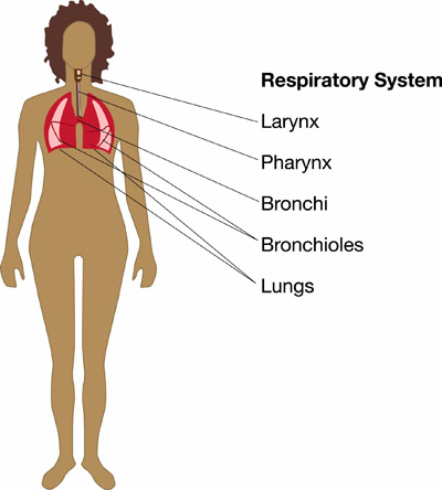
The respiratory system consists of the nose, mouth, pharynx, larynx, trachea, bronchi and lungs. It acts in conjunction with the cardiovascular system to supply oxygen (O2) and remove carbon dioxide (CO2) from the blood.
Cells in the body need O2 for metabolism. They also need to dispose of the by-product CO2. The respiratory and cardiovascular systems work together to provide the mechanism for this to occur. The respiratory system takes in the O2 and disposes of the CO2 and the cardiovascular system pumps both around the body.
The larynx, or voice box, is a passageway that connects the pharynx with the trachea. It contains the thyroid cartilage (commonly called the “Adam’s apple”). At the top of the larynx is the epiglottis, which keeps food from entering the larynx and keeps you from choking.
The trachea is the tube you can feel when you put your hand on the front of your neck. It is reinforced with cartilage rings to prevent it from collapsing. It extends from the larynx and divides into right and left primary (extrapulmonary) bronchi.
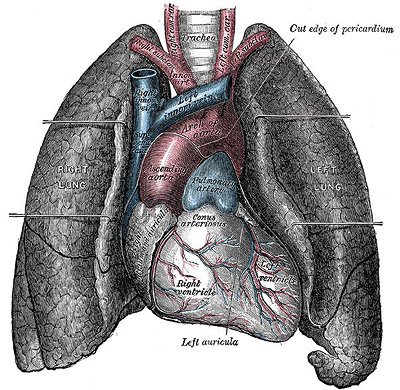
The respiratory bronchioles and the alveoli are where the actual gas exchange takes place, and they always have a little bit of air in them. The lungs are above the diaphragm in the thorax and are enclosed by a pleural membrane called the pulmonary pleura. The parietal pleura lines the thoracic cavity. Pleural fluid keeps everything lubricated so the lungs can move with little friction as you breathe. The right and the left lungs do not look the same. The right lung has three lobes with two fissures. The left lung is smaller, has two lobes with one fissure, and a cardiac notch, which is where the heart nestles.
Air moves in and out of the lungs through pulmonary ventilation: inspiration (inhalation) and expiration (exhalation). This is an autonomic reflex explained by Boyle’s law, which states that the volume of gas (in this case O2 and CO2) varies inversely with pressure. As pressure increases, volume decreases; as pressure decreases, volume increases. These changes in pressure occur with the contraction and relaxation of the diaphragm. When the diaphragm contracts it augments the volume of the lungs and decreases the pressure, when the diaphragm relaxes, the lung volume decreases and pressure increases.
O2 is transported in red blood cells (RBCs) and is bound to hemoglobin (Hb) to form the compound oxyhemoglobin.
There are three ways CO2 is transported in blood:
High levels of CO2 cause blood to become more acidic and this causes Hb to release O2. When O2 enters from the airways and is exchanged in the lungs, it then travels in the blood to the heart where it is pumped to the rest of the body and distributed as needed to cells. As cells pick up the O2 they are also releasing CO2. CO2 hitches a ride on Hb and goes back to the lungs where CO2 is released and exhaled. A rate of flow into and out of the lungs and a high surface area is required for gas exchange to occur. Both O2 and CO2 diffuse from an area of higher partial pressure to a lower internal and external respiration.
The brain has two areas that are part of basic life support. One is the medulla oblongata and the other is the pons. Both of these areas set the rhythm for basic breathing and send messages to the nerves that control the diaphragm. As the diaphragm contracts and relaxes, the process of inspiration and expiration occurs. There are times when the body experiences a chemical imbalance that requires a change in normal breathing. If there is too much CO2 in the body, blood pH drops and it becomes more acidic. The medulla oblongata senses this and modifies the breathing pattern to deep breaths that allow an increase in O2. If there is too much O2 in the body, blood pH increases and becomes more alkaline. The saturated Hb does not allow CO2 to release. The medulla oblongata is alerted and modifies the breathing pattern to shallow breaths. However, if the body continues to take in too much O2, hyperventilation occurs and can result in unconsciousness.
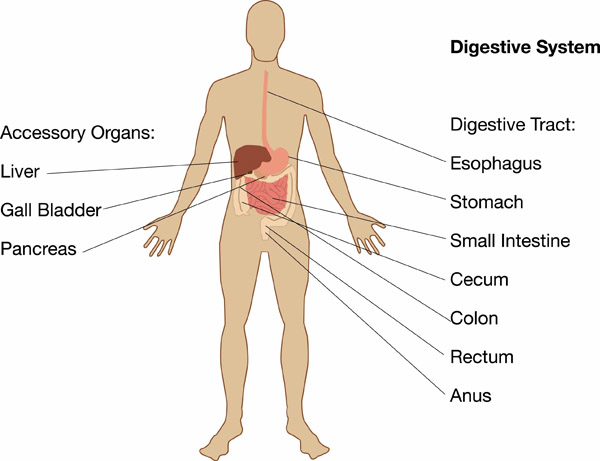
Digestion is the breakdown of food into simpler molecules that can be absorbed by the body. The digestive system is actually a hollow tube called the gastrointestinal tract (GI tract). The first task of the digestive system is to break down the food into a fine pulp. The next task is to chemically act on the food, breaking it down into smaller molecules. The last task is to absorb the molecules and pass them along to the bloodstream for distribution to the rest of the body.
The mouth prepares food for entry into the GI tract. The lips, cheeks, and tongue work together to manipulate food for chewing. The teeth begin the process of mechanical digestion, which is the physical breaking up of food. While the teeth cut and grind food into a pulp, the salivary glands secrete the first digestive enzymes of the GI tract. Saliva has three functions:
As food is swallowed, the tongue moves the food into the pharynx>>, it presses down on the epiglottis. The epiglottis stops entry to the respiratory tract and guides the food to the GI tract.
The food will next pass down the 25cm-wide tube that connects the pharynx to the stomach, the esophagus. The esophagus acts as a kind of descending escalator, moving food down to the stomach. It does this by rhythmic waves of contractions called peristalsis. The esophagus passes through the diaphragm to get to the stomach.
The stomach is a thick muscular sac located just below the diaphragm in the abdomen. It temporarily stores food and mechanically breaks down food. It also chemically unravels and breaks down proteins. Three sets of glands in the stomach lining secrete gastric fluids. The stomach mixes its contents with a churning action. The mixture of food, HCl, and enzymes eventually (after 2 to 3 hours) forms a soupy, semisolid material called chyme.
One set of glands produces mucus, which keeps the food lubricated and protects the walls of the stomach from being digested. Another set of glands secretes hydrochloric acid (HCl), which helps break down food. The release of HCl is regulated by the hormone, gastrin. A third set of glands secretes pepsin, a digestive enzyme that breaks proteins into smaller polypeptides. The pyloric valve between the small intestine and the stomach opens and the food is forced into the small intestine by peristalsis. At this point, most proteins have been broken down, but sugars and fats have not been chemically altered.
The muscle contractions that move food down the esophagus are called
D is correct, because peristalsis is the rhythmic waves of contractions that move food down the esophagus and through the pyloric valve. Chyme is produced in the stomach. Amylase is an enzyme in the mouth. Gastrin is a hormone that regulates the release of HCI in the stomach.
Food passes through the pyloric valve into the small intestine, which is approximately 6m long. The first part of the small intestine is called the duodenum. Here, the chyme is flooded with a variety of enzymes and digestive fluids that further break down the food molecules. The enzymes and digestive enzymes come from three sources: the small intestine, pancreas, and liver.
Glands lining the duodenum release enzymes called peptidases, which continue the process of protein digestion. Other enzymes attach to complex carbohydrates, breaking them down into sugars. When chyme enters the small intestine, glands in the pancreas are stimulated to release pancreatic fluid. The fluid enters a duct that empties into the duodenum. Enzymes in the fluid aid in digestion of carbohydrates, fats, and proteins. The enzymes include amylase, protease, and lipase. Pancreatic fluid also contains sodium bicarbonate, which neutralizes the hydrochloric acid in the chyme.
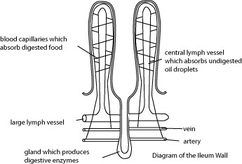
As food enters the small intestine, the liver is stimulated to release bile through the bile duct into the duodenum. Bile is a yellow-brown liquid that is stored in the gall bladder. Bile does not contain enzymes. It is a mixture of cholesterol, colored pigments, and bile salts. Bile salts aid lipase in properly digesting fats. The complete digestion of carbohydrates, fats, and proteins takes place in the duodenum, which is the first 25 cm of the small intestine. The rest of the small intestine is devoted to absorbing water and products of digestion into the bloodstream. The lining of the small intestine is covered with fingerlike projections called villi, which increase the absorptive surface area of the small intestine. The average surface area is about 300 sq meters.
When the food leaves the small intestine, it is essentially nutrient-free. Only water, cellulose, and other indigestible substances are left. The food then passes into the large intestine and colon. The large intestine is about 1m long, but it is about 3 times larger than the small intestine in diameter. Instead of being coiled up, the colon is in three relatively straight segments. The main job of the colon is to remove water from the undigested materials passing through it. The undigested material is then reduced to solid waste products called feces. No digestion takes place in the large intestine. The feces move through the colon by peristalsis and collect at the rectum. At some point, the feces will leave the body through the anus.
It is important to note that many enzymes aid in the digestion process. Here is a short list:

The part of the digestive system in which digested materials are absorbed is the
The correct choice is B. Villi and macrovilli in the small intestine absorb nutrients. The digestive process begins in the mouth, the large intestine removes water from undigested materials. The stomach breaks food particles down for later absorption.
The urinary system is efficient in getting rid of byproducts that are not needed. The kidneys and their associated parts all play a role in making sure that the blood is kept clean and that the body does not lose too much water or other essential nutrients. The urinary system also has other important roles in keeping the balance, or homeostasis of the blood. The organs of the urinary system are the kidneys, ureters, urinary bladder, and the urethra. The kidneys do the bulk of the work, while the other parts act as viaducts or storage for what the kidneys produce.
The kidneys filter many liters of blood every day — approximately one-quarter of the body’s blood flows through the kidneys each minute! In addition, they regulate the blood volume and the chemical makeup of the blood.
The kidneys help regulate blood pressure with the production of an enzyme called rennin. When blood pressure drops, rennin causes blood vessels to constrict, forcing blood pressure to increase. The kidneys produce the hormone erythropoietin, which stimulates the production of red blood cells. The kidneys also convert Vitamin D into a form that is usable to the body.
In addition, the kidneys have four main roles:
The other roles of the kidneys — water and electrolyte balance — are closely linked. Water makes up 50 to 60 percent of body weight depending on gender. Women tend to have more fat than men, and fat has less water than muscle. Two-thirds of this water is found in cells, while the remaining third is found extracellularly in interstitial fluids, like plasma and cerebrospinal fluid.
The re-absorption of water and electrolytes is regulated mostly by two hormones: anti-diuretic hormone (ADH) and aldosterone (controlled by rennin). If there is high blood loss and blood pressure drops, the filtrate drops. Reacting to this change, osmoreceptors in the hypothalamus release ADH. This allows for the absorption of more water in order to increase blood volume and pressure; therefore, only a small amount of concentrated urine is produced. If there is too little Na+ or other ions in the blood, the blood becomes diluted, which causes edema or circulatory collapse. Aldosterone regulates the re-absorption of Na+. As it is reabsorbed, water passively follows Na+.
To review, the kidneys function to regulate these processes:
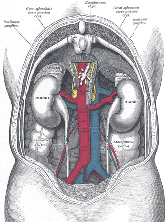
The kidneys are located between T12 and L3 of the spine in the mid- to lower-back and are protected by the ribs. The right kidney is a little lower than the left due to the location of the liver. The indention in the center of the kidney is called the hilus. Kidneys are encased in a protective sheath called the renal capsule. The renal capsule and some adipose tissue hold the kidney against the muscles of the trunk wall.
A kidney opened lengthwise reveals three regions:
The pyramids are arranged with the base of the pyramid toward the cortex and the apex towards the inner part of the kidney. At the end of the pyramids are collection vessels called calyces. The calyces drain into the renal pelvis, a cavity that is continuous with the ureters. The pyramids are separated by renal columns, which are structures that are similar in makeup to the cortex.
Since so much blood passes through the kidneys, they have a great blood supply. The renal artery branches from the abdominal aorta that goes to the kidneys. From there it segments into the segmental artery, to the lobular artery, into the interlobular arteries, and then to the cortex.
The major structural unit of the kidney is the nephron. In each kidney there are millions of nephrons producing urine. A nephron has two main parts: the glomerulus and the renal tubule.
The glomerulus is essentially a capillary bed. The blood goes into the glomerulus through the afferent arteriole and leaves via the efferent arteriole. At one end of the renal tubule there is a capsule (called Bowman’s capsule) that cups around the glomerulus, forming a porous membrane. Extending from the Bowman’s capsule, the renal tubule becomes very convoluted and is surrounded by blood vessels called peritubular capillaries. The renal tubule has three parts: the proximal tubule, the Loop of Henle, and the distal tubule. The proximal tubule ascends down to the Loop of Henle and then ascends back up in the distal tubule.
The major functional part of the kidneys is the
B is the correct answer. The hilus is a depression where vessels, nerves, or ducts enter the kidney. The renal cortex contains the glomeruli and the convoluted tubules. Renal tubules are small structures that filter blood and produce urine.
Filtration occurs between the glomerulus and the Bowman’s capsule under high pressure. This pressure basically pushes everything that is small enough — water, ions, vitamins, amino acids, and wastes — into the Bowman’s capsule. However, if blood pressure is too low, there is not enough pressure and filtration will not occur. As this filtrate runs through the renal tubule, most of it is reabsorbed into the peritubular capillaries.
The peritubular capillaries arise from the efferent arterioles. The capillaries envelop the renal tubules before draining into the venules. The peritubular capillaries are porous and have low pressure, yet have a high absorption of solutes and water during reabsorption. The solutes and water are then returned to the bloodstream.
The body needs water, glucose, amino acids, and ions, so the body reabsorbs almost everything that was pushed out. Reabsorption begins as soon as the filtrate enters the proximal convoluted tubule. Unlike filtration, which was simple passive transport, reabsorption often uses active transport. There are carriers for the substances that the body needs to keep, like glucose and amino acids. These are usually all removed and are not found in urine. However, there are no carriers for the substances not kept — like creatinine and uric acid. Ions are reabsorbed as necessary.
It is while the filtrate is passing through the nephron that urine is formed. Generally, the kidneys filter 150 to180 liters of plasma in 24 hours, yet produce only 1to 2 liters of urine.
Under normal conditions, urine is sterile, yet slightly acidic (~6.0 pH). Coloration ranges from clear to dark yellow, the latter due to the urochrome pigment from the breakdown of hemoglobin. Urine can contain Na+, K+, urea, uric acid, creatinine, ammonia, ions, and vitamins.
Urine needs to leave the body. This is where the other parts of the urinary system come into play.
The ureters are tubes that go from the hilus of the kidney to the urinary bladder. Ureters are smooth muscle and use peristalsis.
The urinary bladder is a smooth, collapsible sac that holds urine. This bladder expands as it fills.
The urethra is a tube that carries the urine from the bladder out of the body. The urethra also operates by peristalsis. It has an internal sphincter that stays closed when urine is not being expelled. Urethra size depends on gender: it is much shorter in females (3 to 4 cm) compared to males (20 cm).