The endocrine system, like the nervous system, is a regulatory system. It is composed of glands that secrete hormones into the bloodstream. Hormones, which are chemical messengers, bind to receptors on cells and affect the behavior of the cells. A response to hormones is generally slower than a response to a nerve impulse. The major hormone-producing organs are the endocrine glands. Endocrine glands do not have ducts; they release their secretions directly into the bloodstream. Endocrine glands are scattered throughout the body and do not have direct connections to one another.

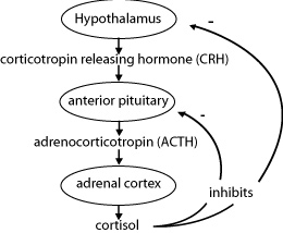
The hypothalamus controls the secretions of the pituitary gland. The activity of the hypothalamus is influenced by the levels of hormones in the blood and sensory information that enters the central nervous system. In the hypothalamus, special neurons extend their axons into the pituitary gland. When the neurons are stimulated, the vesicles at the ends of the axon terminals release their contents into the pituitary gland. The hormones will then be diffused into the capillaries.
The pituitary gland dangles on a slender stalk of tissue at the base of the skull. It is divided into two parts. The front part, the anterior lobe, produces seven major peptide hormones.
The back portion of the pituitary, the posterior lobe, releases two other hormones. ADH (antidiuretic hormone) stimulates the kidneys to reabsorb more water from the collecting tubules that comprise them. Oxytocin stimulates the contraction of muscles in the uterus, which helps a mother to push out a newborn during childbirth. It also causes the release of breast milk of a nursing mother. Oxytocin’s function in males is not known.
The pineal gland is a pea-sized gland located in the top of the brain. It secretes melatonin>>. Other functions of the pineal gland in humans are not yet known. Melatonin seems to be released as a response to darkness, so the pineal gland is thought to be involved in daily biorhythms. It has also been implicated in mood disorders.
The thyroid gland is located at the base of the neck and wraps around the upper part of the trachea just below the larynx. It produces several hormones, but the most important are thyroxine. Thyroxine increases the body’s metabolic rate and promotes the normal growth of the brain, bones, and muscles.
The production of hormones by glands of the endocrine system is initiated by the
A is correct because the hypothalamus acts as the director of the endocrine system by stimulating secretion of hormones by the endocrine glands. The adrenal medulla is a specialized part that secretes neurohormones, adrenaline (or epinephrine), and noradrenaline (norepinephrine). The thyroid gland secretes thyroxine that increases metabolic rate and promotes growth. The pineal gland secretes melatonin in response to darkness.
The parathyroid glands are attached to the back surface of the thyroid gland. There are usually four parathyroid glands, which secrete parathyroid hormone (PTH). PTH regulates the calcium levels in the blood by increasing the reabsorption of calcium in the kidneys and the digestive system. In addition, PTH is important in promoting proper nerve and muscle function, as well as maintaining bone structure.
The adrenal glands are walnut-shaped structures that sit on top of the kidneys, one gland on each kidney. They produce more than two dozen corticosteroids, which are essential for normal body function. One example is aldosterone. Aldosterone regulates the reabsorption of sodium and the excretion of potassium by the kidneys. Cortisol helps to control the rate of metabolism of carbohydrates, fats, and proteins. It also helps people cope with stress and acts to reduce inflammation. In addition, they secrete the neurohormones, such as adrenaline (or epinephrine) and noradrenaline (norepinephrine). These hormones prepare the body for emergency action by increasing heart rate, blood pressure, and blood supply to skeletal muscles. They also increase the conversion of glycogen to glucose and stimulate the body’s metabolic rate. Noradrenaline stimulates the heart muscle.
The pancreas is located just behind the stomach. The hormone-producing part of the pancreas consists of clusters of cells that resemble islands. They are called islets of Langerhans. Each islet secretes insulin and glucagon, which regulate the metabolism of blood glucose. Insulin is a storage hormone that stimulates the ability of its target cells to take up and use glucose. Its major targets are the cells of the liver, skeletal muscles, and fatty tissue. Insulin prevents the level of glucose in blood from rising immediately after a meal. It also ensures that excess glucose will be stored for further use.
The gonads (ovaries and testes) are the body’s reproductive glands. The ovaries produce the female sex hormones, estrogen, and progesterone. Estrogen is required for the development of eggs and for the formation of the physical characteristics of a female. Progesterone prepares the uterus for the arrival of a developing embryo. The testes produce male sex hormones called androgens. Androgens, such as testosterone, are required for normal sperm production and the development of physical characteristics for a male.
Which gland produces cortisol?
The correct choice is D, because cortisol is produced and secreted by the adrenal cortex. The parathyroid secretes parathyroid hormone (PTH). The pancreas secretes insulin and glucagon. The thyroid gland produces several hormones, but most importantly thyroxine.
The endocrine system is regulated by a negative-feedback mechanism. This mechanism functions to maintain homeostasis in the body.
The hormones produced by endocrine glands fall into two groups: polypeptides and steroids. Polypeptides, such as glucagon and thyroxine, are large proteins composed of chains of amino acids. Steroids, such as progesterone, are lipids that are produced from cholesterol. In addition, some cells and tissue produce local hormones called prostaglandins. These hormones are fatty acids.
Secretions from ductless glands are called
The correct choice is C. Endocrine glands are ductless and secrete hormones. Enzymes are proteins that act as catalysts. Excretory fluids come from the urinary system. Digestive fluids come from the digestive system.
For the combination of parental genotypes during the mating process, and a mixture of genetic materials to take place, the sex cells must first go through a process called meiosis, which results in the gametes (eggs and sperm) containing only half of the parental genetic material. Meiosis only occurs in the testes and ovaries to produce gametes.
Meiosis produces haploid gametes via two nuclear divisions: meiosis I and meiosis II. In meiosis I, homologous chromosomes pair up and cross over. The result is two genetically identical daughter cells. In meiosis II, these daughter cells split to form four haploid cells. You should recall that the gametes have a haploid (n) chromosome number, and the somatic (or body) cells have diploid (2n) number of chromosomes.
While the organs used in reproduction are different in males and females, they have complementary components:
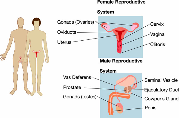
The male reproductive system includes: testes, ductus epididymis, ductus (vas) deferens, ejaculatory duct, urethra, seminal vesicles, prostate gland, bulbourethral glands, and penis. The scrotum, which consists of loose skin and superficial fascia, supports and contains the testes. The testes are paired oval glands that contain seminiferous tubules (where sperm is made), sertoli cells (nourishing cells), and Leydig cells (that produce testosterone). The testes descend into the scrotum through inguinal canals during the seventh month of fetal development. The temperature of the testes is regulated by the contraction of the cremaster and dartos muscles. These muscles bring the testes up or down as needed into the pelvic cavity.
Spermatogenesis is when immature spermatogonia develop into mature sperm. Mature sperm consist of a head, a mid-piece, and a tail. Their function is to fertilize a secondary oocyte. During puberty GnRH stimulates secretion of FSH and LH. LH stimulates the production of testosterone. FSH and testosterone stimulate spermatogenesis.
Testosterone’s role is to control growth and maintenance of sex organs, bone growth, protein anabolism, and sperm maturation. It also stimulates the development of secondary sex characteristics, such as facial hair and deepening of the voice.
The duct system consists of seminiferous tubules, straight tubules and rete testes. Sperm flow out of the testes through efferent ducts. There is also the ductus epididymis, which is a place on the superior portion of the testicle in which the sperm mature. After the sperm mature, they enter the vas deferens, which stores sperm and propels it towards the urethra during ejaculation. Sperm then enter the ejaculatory duct, which is the union of ducts from seminal vesicle and ductus deferens. This duct acts as a passageway way for sperm and secretions of seminal vesicles into the prostatic urethra. There are three parts to the urethra: prostatic, membranous, and spongy (penile).
Accessory sex glands include:
Semen is a mixture of sperm and seminal fluid. It provides transportation and nutrients for the sperm, and it helps to neutralize the acidity of the vagina and male urethra (remember, urine also passes through the male urethra).
The main organs of the female reproductive system include: ovaries, uterine (Fallopian) tubes, uterus, vagina and vulva, and mammary glands.
The ovaries are located in the superior part of the pelvic cavity and are lateral to the uterus. Their job is to produce and discharge secondary oocytes (ovulation is the release of an egg for fertilization), and to secrete estrogens, progesterone, relaxin, and inhibin. An egg is produced through oogenesis, which is the production of haploid secondary oocytes.
The vagina acts as a passageway for menstrual flow, lubricants, and sperm, and serves as the birth canal. External genitalia (vulva) include the mons pubis, labia majora and minora, clitoris, vestibule, vaginal and urethral orifices, hymen, and various glands. The perineum is a diamond-shaped area between the anus and vagina.
Females also have mammary glands, which are modified sweat glands that synthesize, secrete, and release milk, which is regulated by estrogen and progesterone. Milk production is also stimulated by the hormone prolactin, and release is stimulated by the hormone oxytocin.
There are several ways the body protects itself through nonspecific defenses.
Anything that gets through these four nonspecific defense barriers and threatens the body then encounters the immune system. The immune system is the body’s defense against invading microbes and foreign cells, as well as the body’s own cells that have gone bad.
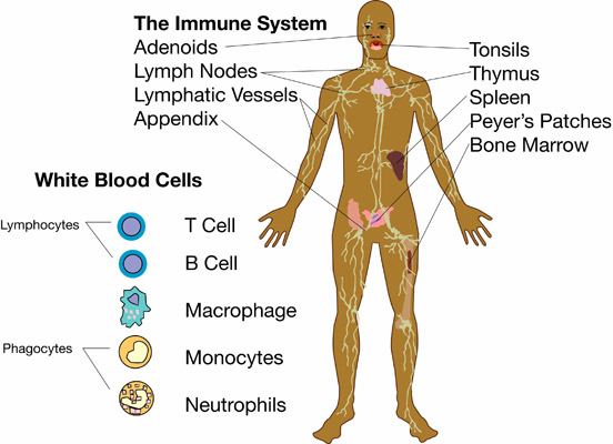
The immune system consists of cells and some molecules. This system has no organs; instead, it uses the pathways of the lymphatic and circulatory systems to transport immune system cells. These cells go hunting for foreign invaders that do not belong in the body. They attack these invaders and disable, kill, or remove them from the body.
The lymphatic system works in conjunction with the immune system. The main function of the lymphatic system is to keep the body healthy. The tubes that carry lymph fluid throughout the body are the lymphatic vessels. These vessels are structured similarly to the circulatory system’s veins. Lymphatic vessels originate as capillaries in spaces between cells. They are found throughout the body, except in avascular tissue, the spleen, bone marrow, or the central nervous system.
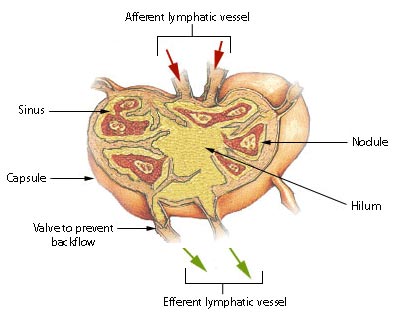
Extra tissue fluid surrounds every cell of the body. Excess tissue fluid is called lymph when it is absorbed by the lymphatic vessels. Lymph moves through the lymphatic system, carrying lymphocytes (white blood cells), a few red blood cells, and fat particles. Lymph fluid is prevented from flowing backward by valves; therefore, the system is a one-way track. The lymph is moved by the squeezing action from the movement of arms, legs, and other skeletal muscles. Lymph passes through lymph nodes where it is filtered to remove microbes and impurities.
The lymph nodes are oval-shaped organs that are located in various places along the lymphatic vessels. These nodes are composed of masses of reticular connective tissue and white blood cells. The nodes act as sort of a net to trap bacteria or other harmful substances. As lymph passes through the nodes, it gets strained and cleaned before being returned to the bloodstream. When the nodes get full, they become enlarged, which is a sign that they are doing their job of cleansing the blood. The lymph nodes are the only organs that filter lymph. Purified lymph returns to the bloodstream via the circulatory system’s veins.
The spleen is a very large lymph node located on the left side of the abdomen, right underneath the diaphragm. It is divided into lobules containing red pulp and white pulp. Red pulp contains red blood cells and white blood cells, including lymphocytes and macrophages. White pulp contains only lymphocytes and macrophages, but no red blood cells.
The spleen serves to filter blood, with the lymphocytes and macrophages filtering out microbes and other foreign material. Excess blood is also stored in the spleen. When the body needs an increase in blood flow, the spleen releases blood.
The thymus gland is located just behind the sternum, overlaying the heart and straddling the trachea. The thymus is large to begin within life and becomes smaller with age. This gland produces the hormone thymosin, which stimulates the differentiation and maturation of T cells.
The tonsils are lymphatic tissue arranged in a ring at the back of the throat. This tissue protects against invasion of inhaled or ingested foreigners. Tonsils “invite infection,” which allows for the production of memory cells for later exposure.
The immune system has a connection with both the skeletal and circulatory systems. White blood cells are created in the red marrow located at the end of long bones, and in the ribs, skull, pelvis, spinal column, clavicles, and sternum. Once created, white blood cells travel throughout the circulatory system.
Specialized cells, called multipotent stem cells, within red bone marrow can potentially differentiate into specific types of blood cells, including white blood cells. There are five types of white blood cells that mount attacks on invaders.
When these cells are stained, the tiny granules in their cytoplasm can be seen easily.
Basophils release histamines. These cells comprise approximately one percent of all white blood cells in the body.
Eosinophils phagocytize antigens and antibodies before splitting them apart. These cells comprise approximately one to four percent of all white blood cells.
Neutrophils phagocytize bacteria. Forty to seventy percent of all white blood cells are neutrophil cells.
The tiny granules in the cytoplasm of agranular leukocytes cannot easily be seen when stained.
Monocytes develop into macrophages. These cells comprise approximately four to eight percent of all white blood cells. Macrophages consume both bacteria and viruses. These cells are involved in the inflammatory, antibody-mediated, and cell-mediated responses. Macrophages play an important role in antigen presentation by engulfing the invader and then putting its antigens on the invaders surface to “present” to T lymphocytes. Macrophages also release proteins that active T cells to release chemicals that will, in turn, transform macrophages into activated macrophages “with a license to kill.”
Approximately 20 to 45 percent of all white blood cells are either B or T lymphocytes. B lymphocytes produce antibodies. T lymphocytes mature in the thymus gland upon stimulation from the thymosin hormone. They destroy cells that contain viruses. There are three types of T cells.
Killer T Cells are also known as cytotoxic T cells. These are involved in the cell-mediated response. They directly attack and kill host cells harboring a virus. Killer T cells remove infected cells in order to protect uninfected cells.
Helper T Cells are involved in both the antibody- and cell-mediated responses. These cells produce a compound called interleukin, which stimulates the rapid division of B cells and killer T cells.
Suppressor T Cells prevent any unnecessary immune responses. These cells come into play when the invader has been destroyed or inactivated. They release chemicals that inhibit the helper T cells, and thereby suppress the formation of antibodies and/or killer T cells.
Sometimes the general attack used in the nonspecific immune response is not enough to stop the spread of an invader. When this happens, the host becomes ill and the immune system kicks into overdrive to protect the body. There are three ways the specific defense system differs from the nonspecific responses.
The specific defense response happens after the host has been exposed to the invader. Once exposed the body creates specific antibodies.
This first-time encounter with an antigen elicits a primary immune response from lymphocytes and their products. There are two types of primary immune responses.
The antibody-mediated immune response targets extracellular organisms, such as bacteria, viruses, fungus, protozoa, and other parasites. In order for an antibody to bind to an antigen, the antigen must be present. An antigen cannot be found if the invader is already in one of the body’s own cells. The following takes place in a bacterial invasion.
All B cells have the same genes for coding the amino acids in the chain but each maturing B cell shuffles the genetic code into one of millions of possible combinations, so the sequence of the amino acids is shuffled. This changes the order of the amino acids, which changes the shape of the protein. Because of this ability, B cells can give rise to a virtually unlimited number of shapes of antibodies. So when a virgin B cell comes into contact with “its” antigen, it really just happened to be there. The virgin B cells are not produced because of the invasion; they are already there. It is our genes that determine which specific foreign substances our immune system will be able to recognize and fight.
The cell-mediated immune response deals with viruses and other pathogens that are already in the host cell (intracellular) and hidden from antibodies. In the cell-mediated immune response, the host cells are killed by killer T cells before the pathogens can replicate and spread to the other cells. The following occurs in a viral infection.
As in the antibody-mediated response, this stimulation of macrophages causes them to produce interleukins, thus stimulating the production of more killer T cells. This again creates a clonal population of killer T-cells — all with the same antigen receptor as the original killer T cell. Some of these clones become memory T cells and become important in the secondary immune response.
When the body rejects an organ transplant or skin graft, killer T cells are one of the reasons why. They recognize the foreign MHC markers on the transplanted organ cells and attack. This is why organ recipients are given immuno-suppressant drugs, which can compromise the body’s attempt to mount immune responses to other pathogens.
The secondary immune response is a response to a previously encountered pathogen. This response can occur in two to three days, is much greater in magnitude than the primary response, and lasts longer. Those B and T cells that were made into memory cells during the primary immune response have been circulating in the body. When a memory cell comes to the same type of antigen that initiated the primary response, it immediately begins dividing and produces a large clonal population within days. Sometimes the attack commences before the host even knows that it was exposed. This is why you catch any particular cold virus only one time. When people say that they are passing the virus around and keep catching it, they are mistaken. They either never recovered from the first bout, they were exposed to different viruses, or they may have a secondary infection from an opportunistic pathogen.
Sounds are just vibrations in the air around us. Deep, low-pitched sounds result from slow vibrations, while faster vibrations cause high-pitched sounds. The ear distinguishes the pitch and volume of sounds.
The external auditory meatus (also known as the ear) is the fleshy part that helps to collect sounds and funnel them into the auditory canal. The auditory canal extends into the bones of the head but stops at the eardrum or tympanum. The eardrum is the beginning of the middle ear. Sound waves strike the eardrum and cause it to vibrate. Three small bones behind the eardrum — the hammer, anvil, and stirrup — transfer the vibration to a fluid-filled chamber within the inner ear. This chamber, which is shaped like a tightly coiled shell, is the cochlea. When the fluid vibrates, tiny hair cells in the cochlea are pushed back and forth, providing stimulation that is turned into nerve impulses. These impulses travel along the auditory nerve to the brain.
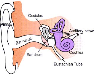
The ears also contain structures for detecting stimuli that make us aware of our movements and allow us to maintain balance. These structures are called semicircular canals.
Each eye is composed of three layers. The outer layer consists of the sclera and the cornea. The sclera consists of tough, white connective tissue. It helps maintain the eye’s shape and provides a place of attachment for the eye muscles. In the front of the eye, the sclera forms a transparent layer, the cornea. Light will first pass through the cornea, which will focus the light onto the back of the eye. The middle layer of the eye consists of the choroid, ciliary body, and the iris. The choroid contains the blood vessels and forms the disk-like iris. The iris gives the eye its color and controls the amount of light entering the eye. In the middle of the iris is a small opening, the pupil, through which light will enter the eye. Just behind the iris is the lens. Small muscles attached to the lens cause it to bend slightly. This bending enables the eye to focus on close and distant objects.
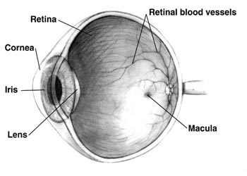
The inner layer of the eye consists of the retina. The retina is the light-sensing portion of the eye. Special photoreceptors are arranged in a layer in the retina. Rods and cones generate nerve impulses, which travel up the optic nerve to the brain. In humans, the field of vision overlaps between the two eyes; each eye sees about one-third of what the other sees. In addition, the two eyes view an object from different angles.
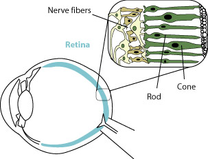
Rods are extremely sensitive to light and can detect various shades of gray, even in dim light. They produce poorly defined images. Cones detect color and are sensitive to edges, so they can detect sharp images.
The outer layer of the eye consists of the
The correct choice is D, the sclera and cornea. Choice A is the middle layer and Choice C is the inner layer. Choice B is part of the ear.
The sense of smell depends on receptors that detect specific chemicals in the environment. In the nose, receptor cells are located in the upper part of the nasal cavity. These receptors contain cilia that extend into the air passageways of the nose and react to chemicals in the air. This reaction causes impulses to travel up the olfactory nerve to the brain.
The sense of taste is also a chemical sense. The organs that detect taste are the taste buds. About 10,000 taste buds are embedded within the surface of the tongue. Taste buds can also be located on the roof of the mouth and on the lips and throat. The taste buds detect four types of chemicals: sugars (sweet), acids (sour), alkaloids (bitter), and metal ions (salty). Generally, the tip of the tongue is sensitive to sweets, while the back of the tongue is sensitive to bitter. The sides of the tongue are sensitive to sour and salty. The taste buds are stimulated when a chemical dissolved in saliva binds to small hairs that protrude from the tip of the taste bud. This generates an impulse that travels to the brain.
The sense of touch is not found in one particular place. All regions of the skin are sensitive to touch. There are several types of sensory receptors that lie just below the surface of the skin.
Sensory receptors for heat and cold are usually scattered. Those for touch are more concentrated in some parts of the body than others. Pain receptors are located throughout the skin.
The largest sense organ is the
The correct choice is C. The skin has sensory receptors covering the body. Choices A, B, and D are all sense organs smaller than the skin.