Over the next four lessons, you will review the structures and functions of the respiratory, urinary, liver, and reproductive systems of the human body.
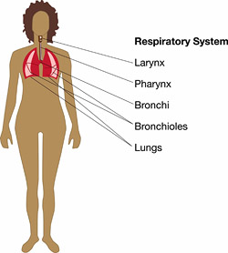
The respiratory system consists of the nose, mouth, pharynx, larynx, trachea, bronchi and lungs. It acts in conjunction with the cardiovascular system to supply oxygen (O2) and remove carbon dioxide (CO2) from the blood.
Cells in the body need O2 for metabolism. They also need to dispose of the by-product CO2. The respiratory and cardiovascular systems work together to provide the mechanism for this to occur. The respiratory system takes in the O2 and disposes of the CO2. The cardiovascular system distributes oxygen to the cells and removes the harmful carbon dioxide.
There are both internal and external organs in the respiratory system . Most of these organs are just conduits to the lungs, which is where the gas exchange actually takes place. Externally, there is the nose and the mouth. The external portion of the nose is made up of cartilage and skin and is lined with a mucous membrane. Opening to the outside are the external nares, or nostrils. The nasal cavity is divided by a septum. The main purpose of the nose, in addition to getting the air into the body, is to moisten, warm, and filter air. It also functions in olfaction and speech.
The nares lead into the internal parts of the nose and mouth. The sinuses are connected to the internal part of the nose. The nasopharynx is also connected to the internal part of the nose.
A tube goes down the throat and splits at the larynx into two tubes: one to the trachea that goes into your lungs and the other to the esophagus that goes into the stomach. The nasopharynx is used only in respiration. As the tube travels farther down toward the stomach and lungs, the name changes according to its placement: oropharynx (near the mouth) and laryngopharynx (near the larynx). Both aid in digestion and respiration.
The larynx, or voice box, is a passageway that connects the pharynx with the trachea . It contains the thyroid cartilage (commonly called “Adam’s apple”). At the top of the larynx is the epiglottis, which keeps food from entering the larynx and keeps you from choking.
The trachea is the tube you can feel when you put your hand on the front of your neck. It is reinforced with cartilage rings to prevent it from collapsing. It extends from the larynx and divides into right and left primary (extrapulmonary) bronchi.
In the lungs, the bronchi branch into the intrapulmonary bronchi. Then, the right bronchus divides into three lobar bronchial branches, while the left divides into two. In the right lung, the lobar bronchi give rise to 10 segmental bronchi; the left lung gives rise to eight segmental bronchi. The branching continues into alveolar ducts, alveolar sacs, and the alveoli.
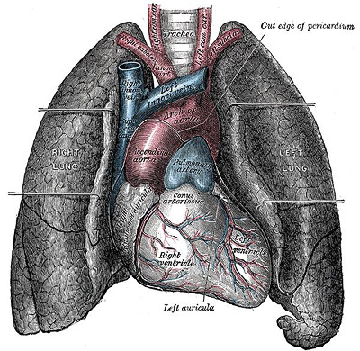
Diagram of Lungs
The respiratory bronchioles and the alveoli are where the actual gas exchange takes place. They always have a little bit of air in them.
The lungs are above the diaphragm in the thoracic and are enclosed by a pleural membrane called the pulmonary pleura. The parietal pleura lines the thoracic cavity. Between these pleural membranes is the pleural fluid that keeps everything lubricated so the lungs can move with little friction as you breathe.
The right and the left lungs do not look the same . The right lung has three lobes with two fissures. The left lung is smaller, has two lobes with one fissure, and a cardiac notch, which is where the heart nestles.
Air gets in and out of the lungs by what is called pulmonary ventilation, and it works on pure physics. Essentially, there is inspiration (inhalation) and expiration (exhalation). This is explained with Boyle’s law, which states that the volume of gas (in this case O2 and CO2) varies inversely with pressure. So as pressure increases, volume decreases; as pressure decreases, volume increases.
This occurs with the contraction and relaxation of the diaphragm. When the diaphragm contracts it pulls the lungs, increases the volume, and decreases the pressure, therefore, air rushes into the lungs. When the diaphragm relaxes, the lung volume decreases and pressure increases, and pushes the air out of the lungs.
O2 is transported in red blood cells (RBCs) and bound to hemoglobin (Hb) to form the compound oxyhemoglobin.
There are three ways CO2 is transported in blood:
High levels of CO2 cause blood to become more acidic and this causes Hb to release its O2. When O2 enters from the airways and is exchanged in the lungs, it then travels in the blood to the heart where it is pumped to the rest of the body and distributed as needed to cells. As cells pick up the O2 they are also releasing CO2. CO2 hitches a ride on Hb and goes back to the lungs where CO2 is released and exhaled.
There is both internal and external gas exchange. Internal gas exchange occurs between capillaries and cells. External exchange occurs between alveoli and pulmonary blood capillaries. A rate of flow into and out of the lungs and a high surface area is required for gas exchange to occur. Both O2 and CO2 diffuse from an area of higher partial pressure to a lower partial pressure via internal and external respiration.
In the brain there are two areas that are part of basic life support. One is the medulla oblongata and the other is the pons. Both of these areas set the rhythm for basic breathing. Normal, relaxed breathing is called eupnea (as opposed to apnea, which is when you stop breathing for a brief moment). Both the medulla oblongata and the pons send messages to the nerves that control the diaphragm. As the diaphragm contracts and relaxes, the process of inspiration and expiration occurs.
There are times when the body experiences a chemical imbalance that requires a change in normal breathing. If there is too much CO2 in the body, blood pH drops and it becomes more acidic. The medulla oblongata senses this and modifies the breathing pattern to slow deep breaths that allow an increase in O2.
If there is too much O2 in the body, blood pH increases and becomes more alkaline. The saturated Hb does not allow CO2 to release. The medulla oblongata is alerted and modifies the breathing pattern to slow, shallow breaths. However, if the body continues to take in too much O2, hyperventilation occurs and can result in unconsciousness. This can be avoided by breathing in and out of a paper bag to decrease O2 intake.